Clinical, radiographic, and histological findings of florid cemento-osseous dysplasia: a case report
- Affiliations
-
- 1Department of Dentistry, Seoul Veterans Hospital, Seoul, Korea. endo95@naver.com
- 2Department of Pathology, Seoul Veterans Hospital, Seoul, Korea.
- KMID: 1449960
- DOI: http://doi.org/10.5624/isd.2011.41.3.139
Abstract
- Cemento-osseous dysplasias are a group of disorders known to originate from periodontal ligament tissue and involve, essentially, the same pathological process. They are usually classified into three main groups: periapical, florid, and focal cemental dysplasias depending on their extent and radiographic appearances. Radiographically, florid cementoosseous dysplasia (FCOD) appears as dense, lobulated masses, often symmetrically located in various regions of the jaws. The best management for the asymptomatic FCOD patient consists of regular recall examinations with prophylaxis. The management of the symptomatic patient is more difficult. A case of FCOD occurring in a 52-year-old edentulous Korean female is reported which is rare with regard to race and sex.
MeSH Terms
Figure
-

Fig. 1 Bilateral buccal bone expansion on posterior quadrant of the maxilla.

Fig. 2 Panoramic radiograph shows diffuse, lobular, irregularly shaped radiopacities or cotton-wool appearance throughout the alveolar process of the quadrants of the edentulous maxilla and mandible.
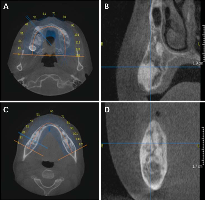
Fig. 3 A. Axial CT image shows high density masses in maxilla premolar-molar regions. B. Coronal CT image show high density masses and their relationship to radiopaque masses and bilateral extensions into the floor of the antrum. C. Axial CT image shows high density masses of the posterior quadrant of the mandible. D. Coronal CT image shows high density masses are surrounded by a low density layer.
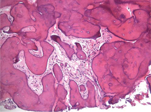
Fig. 4 There are thick, confluent curvilinear trabeculae with little fibrotic stroma. This lesion showed formation of dense sclerotic calcified cementum-like masses. Periphery of the lesions showed globular or ovoid structures of cementoid appearance involved by thin fibrous tissue (H&E stain, ×200).
Cited by 1 articles
-
Recurrent symptomatic cemento-osseous dysplasia: A case report
Chang-Ki Min, Kwang-Joon Koh, Kyoung-A Kim
Imaging Sci Dent. 2018;48(2):131-137. doi: 10.5624/isd.2018.48.2.131.
Reference
-
1. Ong ST, Siar CH. Florid cemento-osseous dysplasia in a young Chinese man. Case report. Aust Dent J. 1997. 42:404–408.
Article2. Miyake M, Nagahata S. Florid cemento-osseous dysplasia. Report of a case. Int J Oral Maxillofac Surg. 1999. 28:56–57.
Article3. Waldron CA. Fibro-osseous lesions of the jaws. J Oral Maxillofac Surg. 1985. 43:249–262.
Article4. Cannon JS, Keller EE, Dahlin DC. Gigantiform cementoma: report of two cases (mother and son). J Oral Surg. 1980. 38:65–70.5. Oikarinen K, Altonen M, Happonen RP. Gigantiform cementoma affecting a Caucasian family. Br J Oral Maxillofac Surg. 1991. 29:194–197.
Article6. Coleman H, Altini M, Kieser J, Nissenbaum M. Familial florid cemento-osseous dysplasia - a case report and review of the literature. J Dent Assoc S Afr. 1996. 51:766–770.7. Toffanin A, Benetti R, Manconi R. Familial florid cementoosseous dysplasia: a case report. J Oral Maxillofac Surg. 2000. 58:1440–1446.
Article8. Gariba-Silva R, Sousa-Neto MD, Carvalho JR Jr, Saquy PC, Pecora JD. Periapical cemental dysplasia: case report. Braz Dent J. 1999. 10:55–57.
Article9. Said-al-Naief NA, Surwillo E. Florid osseous dysplasia of the mandible: report of a case. Compend Contin Educ Dent. 1999. 20:1017–1019.10. Beylouni I, Farge P, Mazoyer JF, Coudert JL. Florid cementoosseous dysplasia: report of a case documented with computed tomography and 3D imaging. Oral Surg Oral Med Oral Pathol Oral Radiol Endod. 1998. 85:707–711.11. Pindborg J, Kramer I, Torloni H. WHO International histological classification of tumors series No.5. Histological typing of odontogenic tumors, jaws cysts and allied lesions. 1971. Geneva: WHO;32–34.12. Melrose RJ, Abrams AM, Mills BG. Florid osseous dysplasia. A clinical-pathologic study of thirty-four cases. Oral Surg Oral Med Oral Pathol. 1976. 41:62–82.13. Oikarinen K, Altonen M, Happonen RP. Gigantiform cementoma affecting a Caucasian family. Br J Oral Maxillofac Surg. 1991. 29:194–197.
Article14. Young SK, Markowitz NR, Sullivan S, Seale TW, Hirschi R. Familial gigantiform cementoma: classification and presentation of a large pedigree. Oral Surg Oral Med Oral Pathol. 1989. 68:740–747.
Article15. MacDonald-Jankowski DS. Florid cemento-osseous dysplasia: a systematic review. Dentomaxillofac Radiol. 2003. 32:141–149.
Article16. Schneider LC, Mesa ML. Differences between florid osseous dysplasia and chronic diffuse sclerosing osteomyelitis. Oral Surg Oral Med Oral Pathol. 1990. 70:308–312.
Article
- Full Text Links
-
- Actions
-
Cited
- CITED
-
- Close
- Share
-
- Similar articles
-
- 3 Types of Cemento-Osseous Dysplasia: Case Reports
- Misdiagnosis of florid cemento-osseous dysplasia leading to unnecessary root canal treatment: a case report
- Florid cemento-osseous dysplasia: a report of two cases
- Recurrent symptomatic cemento-osseous dysplasia: A case report
- Bisphosphonate-Related Osteonecrosis in a Patient with Florid Cemento-Osseous Dysplasia
