Anat Cell Biol.
2011 Sep;44(3):241-243. 10.5115/acb.2011.44.3.241.
Bilateral variations of the head of the digastric muscle in Korean: a case report
- Affiliations
-
- 1Medical Course, Keimyung University School of Medicine, Daegu, Korea.
- 2Department of Anatomy, Keimyung University School of Medicine, Daegu, Korea. ijchoi@dsmc.or.kr
- 3Institute for Medical Genetics, Keimyung University School of Medicine, Daegu, Korea.
- 4Hanvit Institute for Medical Genetics, Daegu, Korea.
- KMID: 1447437
- DOI: http://doi.org/10.5115/acb.2011.44.3.241
Abstract
- The digastric muscle, as the landmark in head and neck surgery, has two bellies, of which various variations have been reported. In the submental region of a 72-year-old Korean male cadaver, bilateral variations were found in the anterior belly of the digastric muscle. Two accessory bellies, medial to the two normal anterior bellies of the digastric muscle, ran posterior and medially, merging and attaching at the mylohyoid raphe of the mylohyoid muscle. The 3rd accessory belly originated from the right intermediate tendon and ran horizontally, merging the right lower bundle of the right accessory belly and inserted together. These accessory bellies had no connection with the left anterior belly. This unique variation has not been reported in the literature previously, and this presentation will guide clinicians during surgical interventions and radiological diagnoses.
Keyword
Figure
-
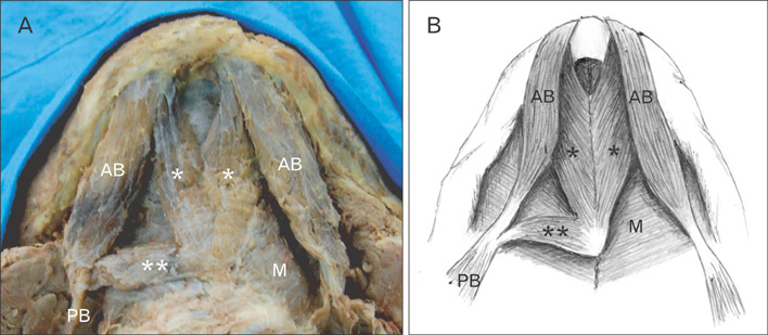
Fig. 1 Photograph (A) and schematic drawing (B) showing the accessory bellies of the digastric muscle. Two accessory bellies (*) merged and some parts are attached to the mylohyoid raphe. Right inferior portion continued as 3rd accessory belly (**) and inserted right intermediate tendon. AB, anterior belly; M, mylohyoid muscle; PB, posterior belly.
Cited by 1 articles
-
A clinical perspective on the anatomical study of digastric muscle
Nandini Prashanth Bhat, Suhani Sumalatha, Ashwija Shetty, Sushma Prabhath
Anat Cell Biol. 2023;56(4):441-447. doi: 10.5115/acb.23.043.
Reference
-
1. Traini M. Bilateral accessory digastric muscles. Anat Clin. 1983. 5:199–200.2. Ziółkowski M, Marek J, Kłak A. The human digastric muscle in the fetal period. Folia Morphol (Warsz). 1984. 43:243–249.3. Larsson SG, Lufkin RB. Anomalies of digastric muscles: CT and MR demonstration. J Comput Assist Tomogr. 1987. 11:422–425.4. Fujimura A, Onodera M, Feng XY, Osawa T, Nara E, Nagato S, Matsumoto Y, Sasaki N, Nozaka Y. Abnormal anterior belly of the digastric muscle: a proposal for the classification of abnormalities. Anat Sci Int. 2003. 78:185–188.5. Kim DH, Do HJ, Kim HJ, Won SY, Choi DY, Hu KS, Choi JH, Kim HJ. Anatomic variation of the anterior belly of digastric muscle and positional relationship between the posterior belly of digastric and stylohyoid muscle. Korean J Phys Anthropol. 2010. 23:9–16.6. Celik H, Yilmaz E, Atasever A, Durgun B, Taner D. Bilateral anatomical anomaly of anterior bellies of digastric muscles. Kaibogaku Zasshi. 1992. 67:650–651.7. Sargon MF, Celik HH. An abnormal digastric muscle with three bellies. Surg Radiol Anat. 1994. 16:215–216.8. Uslu SS, Atilla S, Celik HH, Inal E. An important anatomic variation in head and neck region: anomaly of the anterior belly of the digastric muscle. Bull Assoc Anat (Nancy). 1995. 79:39–41.9. Ozgur Z, Govsa F, Ozgur T. The cause of the difference in the submental region: aberrant muscle bundles of the anterior belly of the digastric muscle. J Craniofac Surg. 2007. 18:875–881.10. Aktekin M, Kurtoğlu Z, Oztürk AH. A bilateral and symmetrical variation of the anterior belly of the digastric muscle. Acta Med Okayama. 2003. 57:205–207.
- Full Text Links
-
- Actions
-
Cited
- CITED
-
- Close
- Share
-
- Similar articles
-
- Accessory Heads of Anterior Belly of Digastric Muscle in Korea
- Anatomy and variations of digastric muscle
- A clinical perspective on the anatomical study of digastric muscle
- Anatomic Variation of the Anterior Belly of Digastric Muscle and Positional Relationship between the Posterior Belly of Digastric and Stylohyoid Muscle
- Correction of post-traumatic anterior open bite by injection of botulinum toxin type A into the anterior belly of the digastric muscle: case report
