Rapidly Progressing Germinoma Arising in the Lateral Ventricle and This Mimicked Choroid Plexitis
- Affiliations
-
- 1Department of Radiology, Chung-Ang University College of Medicine, Korea. flightdr61@cau.ac.kr
- 2Department of Neurology, Chung-Ang University College of Medicine, Korea.
- 3Department of Neurosurgery, Chung-Ang University College of Medicine, Korea.
- 4Department of Pathology, Chung-Ang University College of Medicine, Korea.
- KMID: 1443576
- DOI: http://doi.org/10.3348/jksr.2011.64.1.11
Abstract
- There are no previous reports of germinoma primary arising in the lateral ventricle. We describe here a case of a 50-year-old man with lateral ventricular germinoma that showed rapid progression, and this tumor mimicked choroid plexitis.
Figure
-
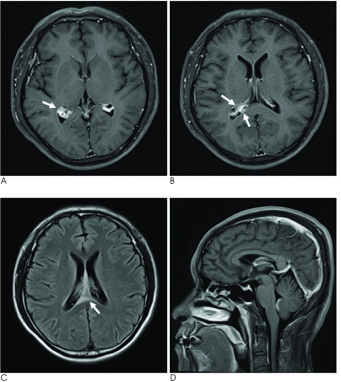
Fig. 1 The axial postcontrast T1-weighted MR image (A and B) demonstrated the enlarged choroid plexus (arrow in figure A) and the adjacent ependymal enhancement in the trigone of the right lateral ventricle (arrows in figure B), and there is suspicious ependymal enhancement and a mildly enlarged choroid plexus in the left lateral ventricle. The axial FLAIR MR image (C) showed high signal intensities in the septum pellucidum (arrow in figure C) and along the trigone of the right lateral ventricle. The sagittal postcontrast T1-weighted MR image (D) demonstrated a suspicious mildly enlarged choroid plexus in the fourth ventricle, and no abnormal lesion in the pineal or suprasellar regions.
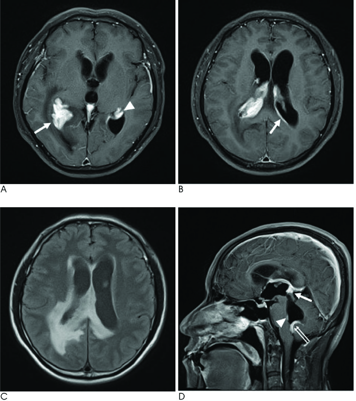
Fig. 2 The axial postcontrast T1-weighted MR images (A and B) demonstrate more progression of the dense enhancement of the enlarged choroid plexus (arrow in figure A) and adjacent ependymal enhancement in the right lateral ventricle and obvious dense enhancement of the enlarged choroid plexus (arrowhead in figure A) and ependymal enhancement (arrow in figure B) in the left ventricle. There are newly enhancing lesions in the ependyma of the right lateral ventricle. The axial FLAIR MR image (C) showed progression of the high signal intensities around both the lateral ventricles, and especially along the right lateral ventricle. The sagittal postcontrast T1-weighted MR image (D) depicts definite dense enhancement of the enlarged choroid plexus in the fourth ventricle (double arrows in figure D) and newly enhancing lesions have appeared in the pineal region (arrow in figure D) and the anterior wall of the fourth ventricle (arrowhead in figure D).
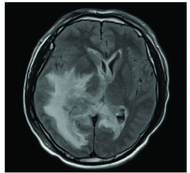
Fig. 3 Axial FLAIR MRI showed extensive white matter edema around both lateral ventricles, and especially along the right lateral ventricle, and there is midline shifting to the left.

Fig. 4 The hematoxylin and eosin stained section shows primitive germ cells with an infiltration of small lymphoid cells (200 ×). Giant cells are noted among the tumor cells (arrows, the figure at the right lower portion [400 ×]).
Reference
-
1. Tamaki N, Lin T, Shirataki K, Hosoda K, Kurata H, Matsumoto S, et al. Germ cell tumors of the thalamus and the basal ganglia. Childs Nerv Syst. 1990; 6:3–7.2. Higano S, Takahashi S, Ishii K, Matsumoto K, Ikeda H, Sakamoto K. Germinoma originating in the basal ganglia and thalamus: MR and CT evaluation. AJNR Am J Neuroradiol. 1994; 15:1435–1441.3. Sartori S, Laverda AM, Calderone M, Carollo C, Viscardi E, Faggin R, et al. Germinoma with synchronous involvement of midline and off-midline structures associated with progressive hemiparesis and hemiatrophy in a young adult. Childs Nerv Syst. 2007; 23:1341–1345.4. Hoque R, Menon U, Gonzalez-Toledo E, Gu X, Jaffe SL. CNS germinoma of the pituitary and pineal regions, lateral ventricle, and fourth ventricle presenting in adulthood. J La State Med Soc. 2008; 160:319–321.5. Smirniotopoulos JG, Rushing EJ, Mena H. Pineal region masses: differential diagnosis. Radiographics. 1992; 12:577–596.6. Kon H, Kumabe T, Jokura H, Shirane R. Recurrent intracranial germinoma outside the initial radiation field with progressive malignant transformation. Acta Neurochir. 2002; 144:611–616.7. Kobayashi T, Kageyama N, Kida Y, Yoshida J, Shibuya N, Okamura K. Unilateral germinomas involving the basal ganglia and thalamus. J Neurosurg. 1981; 55:55–62.8. Tekkok IH, Sav A. Aggressive spinal germinoma with ascending metastases. J Neurooncol. 2005; 75:135–141.9. Naeini RM, Yoo JH, Hunter JV. Spectrum of choroid plexus lesions in children. AJR Am J Roentgenol. 2009; 192:32–40.10. Cho IC, Chang KH, Kim YH, Kim SH, Yu IK, Han MH. MRI features of choroid plexitis. Neuroradiology. 1998; 40:303–307.
- Full Text Links
-
- Actions
-
Cited
- CITED
-
- Close
- Share
-
- Similar articles
-
- A case of entrapped temporal horn of lateral ventricle caused by Pseudomonas stutzeri choroid plexitis
- Choroid Plexus Papilloma in Infant
- A Case of Lateral Ventricle Choroid Plexus Papilloma in a Child
- A Case of Mixed Germ Cell Tumor in the Third and Lateral Ventricle
- A Case of Lateral Ventricle Choroid Plexus Papilloma in an Infant
