Candida Parapsilosis Arthritis Involving the Ankle in a Diabetes Patient: A Case Report1
- Affiliations
-
- 1Department of Radiology, The Catholic University of Korea Uijeongbu St. Mary's Hospital, Korea. ka1000@catholic.ac.kr
- KMID: 1443509
- DOI: http://doi.org/10.3348/jksr.2011.64.6.587
Abstract
- Candida parapsilosis is a rare opportunistic fungal pathogen of the musculoskeletal region. Immune function of almost all patients is severely disturbed. Most reported cases of septic arthritis of joints by Candida involve the knee, especially Candida parapsilosis. To our knowledge, there has been only one case report of Candida parapsilosis involving the ankle presented on only plain radiography. We report a case of Candida parapsilosis arthritis involving the ankle in a diabetes patient which was shown on MR imaging.
MeSH Terms
Figure
-
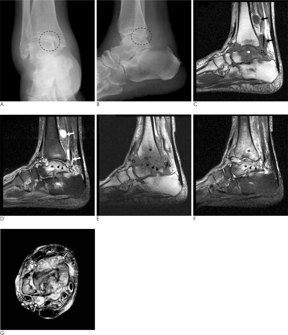
Fig. 1 63-year-old woman with candida arthritis. A, B. Anteroposterior (A) and lateral (B) radiographs of the right ankle show asymmetric joint space narrowing and subchondral bone erosion at the posterior and medial aspect of tibiotalar joint (dotted circle). Moderate swelling of soft tissue around the ankle joint is seen. C, D. Sagittal T1-(C) and fat-supressed T2-weighted (D) MR images show joint effusion and synovial thickening. Two loose (rice) bodies (open arrows) are seen within the distended joint space, with iso to slightly high signal intensity on T1-weighted image and low signal intensity on T2-weighted image. Distension of the flexor hallucis longus and tibialis posterior tendon sheaths by fluid collection (solid arrows) represents combined tenosynovitis. Subchondral bone erosion was seen at the posterior aspect of the talar dome (arrowheads). Bone marrow edema (asterisks) is noted in the talus as a low and high signal intensity on T1-and T2-weighted images, respectively. E, F. Four months later, follow-up sagittal T1- (E) and fat suppressed T2-weighted (F) MR images show increased joint effusion and rice bodies within the joint space (open arrows). Destruction of the talar dome by complicated subchondral erosion (arrow heads) is aggravated. Hyperintense bone marrow signals in tibia and talus (asterisks) also progressed, representing concomitant osteomyelitis. G. Axial fat suppressed T2-weighted image at the tibiotalar joint level more clearly reveals abudant hypointense rice bodies (arrows) and a moderate amount of joint effusion.

Fig. 2 Photomicrographs of the patients A. Photomicrograph shows chronic granulomatous inflammtion with multinucleated giant cells (hematoxylin and-eosin, magnification ×40). B, C. Photomicrographs for PAS (B) and methenamine-silver (C) staining show several yeast-forming fungal organisms (arrow) (×400).
Reference
-
1. Masoud M, Nasser NJ, Karban A, Edelstein S. Candida parapsilosis septic arthritis in a renal transplant patient. J Clin Rheumatol. 2008; 14:56.2. Marmor L, Peter JB. Candida arthritis of the knee joint. Clin Orthop Relat Res. 1976; 133–135.3. Turgut B, Vural O, Demir M, Kaldir M. Candida arthritis in a patient with chronic myelogenous leukemia (CML) in blastic transformation, unresponsive to fluconazole, but treated effectively with liposomal amphotericin B. Ann Hematol. 2002; 81:529–531.4. Trofa D, Gacser A, Nosanchuk JD. Candida parapsilosis, an emerging fungal pathogen. Clin Microbiol Rev. 2008; 21:606–625.5. Miller DJ, Mejicano GC. Vertebral osteomyelitis due to Candida species: case report and literature review. Clin Infect Dis. 2001; 33:523–530.6. Cha JG, Hong HS, Koh YW, Kim HK, Park JM. Candida albicans osteomyelitis of the cervical spine. Skeletal Radiol. 2008; 37:347–350.7. Munk PL, Lee MJ, Poon PY, O'Connell JX, Coupland DB, Janzen DL, et al. Candida osteomyelitis and disc space infection of the lumbar spine. Skeletal Radiol. 1997; 26:42–46.8. Hsu CY, Lu HC, Shih TT. Tuberculous infection of the wrist: MRI features. AJR Am J Roentgenol. 2004; 183:623–628.9. Parmar H, Shah J, Patkar D, Singrakhia M, Patankar T, Hutchinson C. Tuberculous arthritis of the appendicular skeleton: MR imaging appearances. Eur J Radiol. 2004; 52:300–309.10. Karchevsky M, Schweitzer ME, Morrison WB, Parellada JA. MRI findings of septic arthritis and associated osteomyelitis in adults. AJR Am J Roentgenol. 2004; 182:119–122.
- Full Text Links
-
- Actions
-
Cited
- CITED
-
- Close
- Share
-
- Similar articles
-
- A Case of Candida Parapsilosis Ankle Arthritis after Intra-articular Steroid Injection
- A Case of Candida parapsilosis Arthritis
- A Case of Candida Parapsilosis Infectious Arthritis in a Patient with Enteropathic Arthritis and Ulcerative Colitis
- A Case of Melanonychia Caused by Candida parapsilosis
- A Case of Candida parapsilosis Meningitis in a Patient with Ventriculo-peritoneal Shunt
