Gastrointestinal Stromal Tumor of the Appendix Mimicking a Mucinous Cystadenocarcinoma: A Case Report
- Affiliations
-
- 1Department of Radiology, Bucheon Hospital, Soonchunhyang University College of Medicine, Bucheon, Korea. hklee@schmc.ac.kr
- 2Department of Pathology, Bucheon Hospital, Soonchunhyang University College of Medicine, Bucheon, Korea.
- KMID: 1443497
- DOI: http://doi.org/10.3348/jksr.2011.65.1.81
Abstract
- A gastrointestinal stromal tumor of the appendix is a rare entity. Only a few cases have been reported in this location to date. We present here a case of a pathologically confirmed gastrointestinal stromal tumor of the appendix mimicking a mucinous cystadenocarcinoma in a 67-year-old man.
MeSH Terms
Figure
-
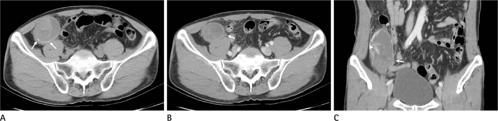
Fig. 1 A. Pre-contrast axial CT image shows a cystic mass with internal soft-tissue component in the right lower quadrant abdominal cavity, which appeared to abut to the cecum in the other CT images. Note the wall calcification (arrows). B, C. Post-contrast axial (B) and coronal (C) CT images show the same cystic mass with irregular, enhancing wall thickening. The internal soft-tissue component (arrowhead) enhances heterogeneously. Note the tubular structure (arrow) abutting the mass, which was connected with the mass in other CT images, representing that the mass arises from the appendix.
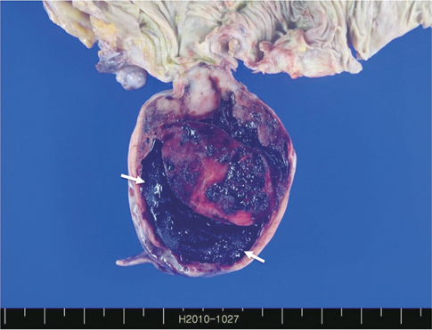
Fig. 2 The cut surface of the surgical specimen shows a tan-white mass with extensive areas of hemorrhage and cystic degeneration (arrows).
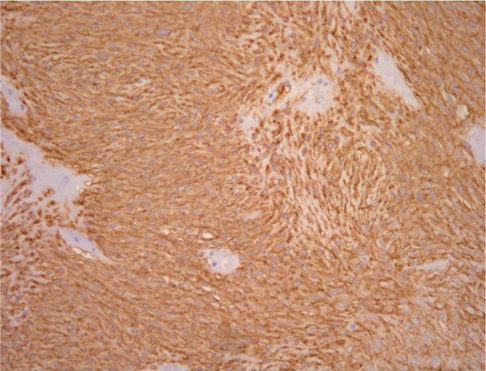
Fig. 3 Photomicrograph of histopathologic specimen shows that the tumor cells are strongly reactive for C-Kit in immunohistochemical stain (× 200).
Reference
-
1. Pickhardt PJ, Levy AD, Rohrmann CA Jr, Kende AI. Primary neoplasms of the appendix: radiologic spectrum of disease with pathologic correlation. Radiographics. 2003; 23:645–662.2. Miettinen M, Lasota J. Gastrointestinal stromal tumors: pathology and prognosis at different sites. Semin Diagn Pathol. 2006; 23:70–83.3. Miettinen M, Sobin LH. Gastrointestinal stromal tumors in the appendix: a clinicopathologic and immunohistochemical study of four cases. Am J Surg Pathol. 2001; 25:1433–1437.4. Yap WM, Tan HW, Goh SG, Chuah KL. Appendiceal gastrointestinal stromal tumor. Am J Surg Pathol. 2005; 29:1545–1547.5. Kim KJ, Moon W, Park MI, Park SJ, Lee SH, Chun BK. Gastrointestinal stromal tumor of appendix incidentally diagnosed by appendiceal hemorrhage. World J Gastroenterol. 2007; 13:3265–3267.6. Elazary R, Schlager A, Khalaileh A, Appelbaum L, Bala M, Abu-Gazala M, et al. Malignant appendiceal GIST: case report and review of the literature. J Gastrointest Cancer. 2010; 41:9–12.7. Badalamenti G, Rodolico V, Fulfaro F, Cascio S, Cipolla C, Cicero G, et al. Gastrointestinal stromal tumors (GISTs): focus on histopathological diagnosis and biomolecular features. Ann Oncol. 2007; 18:Suppl 6. vi136–vi140.8. Miettinen M, Lasota J. Gastrointestinal stromal tumors--definition, clinical, histological, immunohistochemical, and molecular genetic features and differential diagnosis. Virchows Arch. 2001; 438:1–12.9. Levy AD, Remotti HE, Thompson WM, Sobin LH, Miettinen M. Gastrointestinal stromal tumors: radiologic features with pathologic correlation. Radiographics. 2003; 23:283–304. 456quiz 532.10. Gore R, Levine M. Textbook of gastrointestinal imaging. 3rd ed. Philadelphia: Saunders;2008. p. 593–648.
- Full Text Links
-
- Actions
-
Cited
- CITED
-
- Close
- Share
-
- Similar articles
-
- A Case of Sarcomatoid Carcinoma Arising from Mucinous Cystadenocarcinoma of Appendix
- Mucinous Tumors of the Appendix Associated with Mucinous Tumors of the Ovary and Pseudomyxoma Peritonei: A Clinicopathologic Analysis of 5 Cases Supporting an Appendiceal Origin
- A Case of Appendiceal Mucocele found during Total Hysterectomy
- Each Case of Benign and Malignant Mucocele of the Appendix
- Appendiceal Intussusception Caused by Mucinous Cystadenocarcinoma
