FDG-PET/CT Complements Bone Scan with Respect to the Detection of Skip Metastasis of Osteosarcoma: A Case Report
- Affiliations
-
- 1Department of Orthopedic Surgery, Korea University Hospital, Seoul, Korea. pjh1964@hanmail.net
- 2Department of Nuclear Medicine, Korea University Hospital, Seoul, Korea.
- KMID: 1438413
- DOI: http://doi.org/10.5292/jkbjts.2012.18.1.45
Abstract
- Skip lesion is not uncommon feature in osteosarcoma and considered to be importantly associated with poor prognosis factor, and thus, should be excised with the main mass. The accurate pre-operative evaluation of the intramedullary extent of osteosarcoma is essential, because it determines the level of bone resection. Among the reliable detection methods, bone scan has a drawback of high rate of false negative results and regional MRI has a difficulty to cover the whole involved lesions without clinical suspicion. The authors report a case of osteosarcoma of the distal femur with a proximal skip lesion that was not detected by either regional MR imaging or by bone scan, but which was visualized by FDG-PET/CT.
MeSH Terms
Figure
-
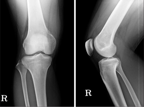
Figure 1 Plain radiographs show an osteolytic lesion of a medial aspect of distal femur in AP view.
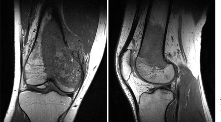
Figure 2 Coronal and sagittal T1-weighted MR image at presentation show an osteosarcoma extending approximately 10 cm from the distal femoral articular surface. There is some soft tissue extension in medial direction.

Figure 3 Whole body bone scan at presentation showing isolated area of increased uptake in medial aspect of right distal femur. There is no suspected metastatic and skip lesion.
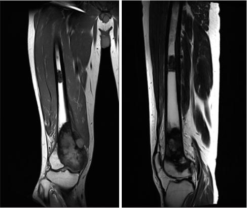
Figure 4 MR imaging of Right femur at the time of pre-operative restaging examination. T1 coronal and sagittal image of whole femur showing the skip lesion in proximal diaphysis.
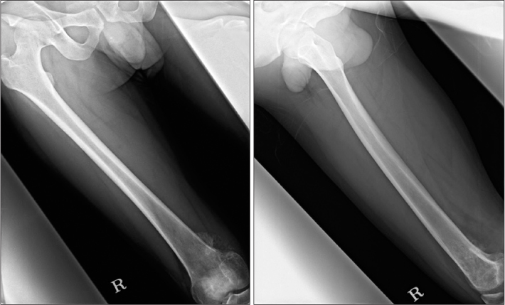
Figure 5 Plain femur AP and Lat view showing no significant skip lesion of the proximal diaphysis.
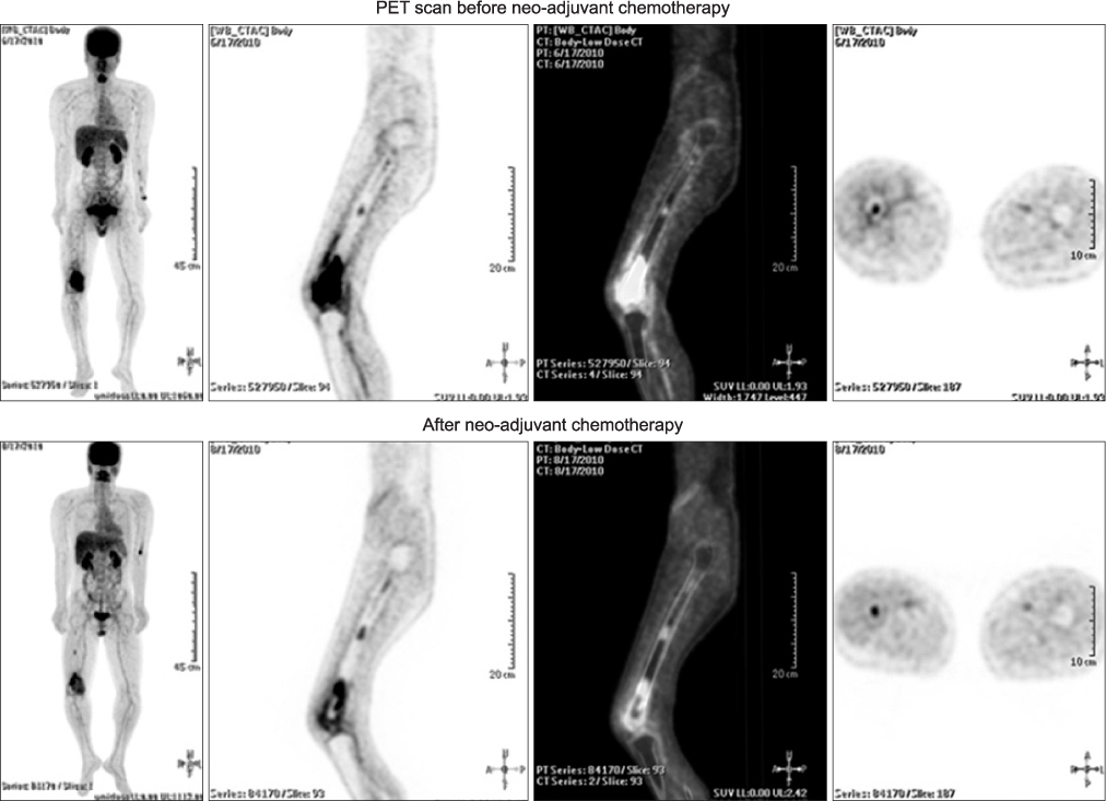
Figure 6 Initial FDG-PET scan and restaging PET scan also showing distinct skip lesion.
Reference
-
1. Enneking WF, Kagan A. "Skip" metastases in osteosarcoma. Cancer. 1975. 36:2192–2205.2. Enneking WF, Kagan A. The implications of "skip" metastases in osteosarcoma. Clin Orthop Relat Res. 1975. (111):33–41.3. Wuisman P, Enneking WF. Prognosis for patients who have osteosarcoma with skip metastasis. J Bone Joint Surg Am. 1990. 72:60–68.4. Cheon GJ, Chung JK, Kim YK, et al. Comparasion of whole body F-18 FDG PET and Tc-99m MDP bone scan for the assessment of metastatic bone lesions. World J Nucl Med. 2003. 2:18–29.5. Gosheger G, Gebert C, Ahrens H, Streitbuerger A, Winkelmann W, Hardes J. Endoprosthetic reconstruction in 250 patients with sarcoma. Clin Orthop Relat Res. 2006. 450:164–171.6. Grimer RJ. Surgical options for children with osteosarcoma. Lancet Oncol. 2005. 6:85–92.7. Ardran GM. Bone destruction not demonstrable by radiography. Br J Radiol. 1951. 24:107–109.8. Berquist TH. Magnetic resonance imaging of primary skeletal neoplasms. Radiol Clin North Am. 1993. 31:411–424.9. Basu S, Alavi A. Bone marrow and not bone is the primary site for skeletal metastasis: critical role of [18F]fluorodeoxyglucose positron emission tomography in this setting. J Clin Oncol. 2007. 25:1297. author reply 1297-9.10. Hur J, Yoon CS, Ryu YH, Yun MJ, Suh JS. Accuracy of fluorodeoxyglucose-positron emission tomography for diagnosis of single bone metastasis: comparison with bone scintigraphy. J Comput Assist Tomogr. 2007. 31:812–819.
- Full Text Links
-
- Actions
-
Cited
- CITED
-
- Close
- Share
-
- Similar articles
-
- Value of Bone Scan in Addition to F-18 FDG PET/CT and Characteristics of Discordant Lesions between F-18 FDG PET/CT and Bone Scan in the Spinal Bony Metastasis
- Discrepancy of Bone Metastases between F-18 FDG PET/CT and Bone Scan in a Patient with Prostate Cancer
- â¶â¸Gallium-Arginine-Glycine-Aspartic Acid and ¹â¸F-Fluorodeoxyglucose Positron Emission Tomography/Computed Tomography in Chondroblastic Osteosarcoma of the Skull
- Case Report of Prostate Cancer Patient with Only Lymph Node Involvement on F-18 FDG PET/CT
- Use of 18F-FDG PET/CT in Second Primary Cancer
