Treatment Strategy for Parkinsonian Diseases Through Mesenchymal Stem Cells
- Affiliations
-
- 1Department of Neurology and Brain Research Institute, Yonsei University College of Medicine, Severance Biomedical Science Institute, Yonsei University, Seoul, Korea. phisland@chol.net
- KMID: 1436766
- DOI: http://doi.org/10.7599/hmr.2012.32.3.145
Abstract
- Parkinsonian diseases including Parkinson's disease (PD) and multiple system atrophy (MSA) are neurodegenerative diseases representative of alpha-synucleinopathies characterized pathologically by alpha-synuclein-abundant Lewy bodies and glial cytoplasmic inclusions, respectively. Cell therapy using mesenchymal stem cells (MSCs) is attractive clinically because these cells are free from ethical and immunological problems. MSCs are present in adult bone marrow and represent <0.01% of all nucleated bone marrow cells. MSCs are multipotent, and differentiation under appropriate conditions into chondrocytes, skeletal myocytes, and neurons has been demonstrated thus far. According to recent studies, the neuroprotective effect of MSCs is mediated by the production of various trophic factors that contribute to functional recovery, neuronal cell survival, and endogenous regeneration of neural tissues. Additionally, MSCs appear to have immunoregulatory properties that can ameliorate the progression of disease. However, the therapeutic use of MSCs as neuroprotectives in PD and MSA has seldom been studied. Here we comprehensively review recent advances in clinical strategies using MSCs in PD and MSA, especially focusing on their neuroprotective properties in preventing or delaying disease progression and therapeutic potential for providing functional recovery.
MeSH Terms
-
Adult
Bone Marrow
Bone Marrow Cells
Cell Survival
Chondrocytes
Disease Progression
Humans
Inclusion Bodies
Lewy Bodies
Mesenchymal Stromal Cells
Multiple System Atrophy
Muscle Fibers, Skeletal
Neurodegenerative Diseases
Neurons
Neuroprotective Agents
Parkinson Disease
Parkinsonian Disorders
Regeneration
Tissue Therapy
Neuroprotective Agents
Figure
-
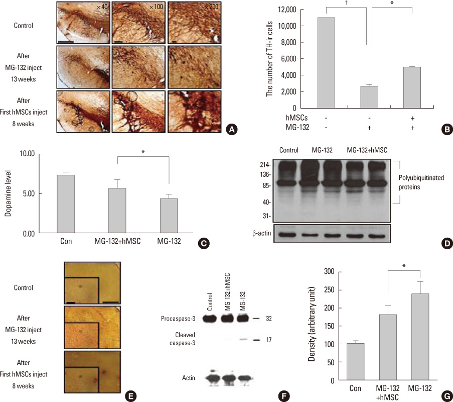
Fig. 1 Neuroprotective effects of human mesenchymal stem cells (hMSCs) in animal model of Parkinson's disease treated with MG-132. Immunohistochemical analysis showed that hMSC treatment dramatically reduced the decline in the number of TH-ir cells in the SN of MG-132-treated rats (A). Stereological analysis revealed that the number of TH-ir cells was significantly higher in the hMSC-treatment group than in the group treated with MG-132 alone (n=5; p<0.05, B). Dopamine levels in the striatum (as assessed by gas chromatography-mass spectrometry) were significantly lower in MG-132-treated rats than in controls (p<0.01); however, hMSC treatment significantly increased the dopamine level in the striatum of MG-132-treated rats (n=5; p<0.05, C). MG-132 treatment resulted in the accumulation of polyubiquitinated proteins and a marked increase in OX-6 immunoreactivity; however, hMSC treatment markedly decreased the accumulation of polyubiquitinated proteins and OX-6 immunoreactivity in MG-132-treated rats (D and E). The level of the cleaved form of caspase-3 was significantly lower in rats treated with hMSCs (F) than in MG-132-treated rats (n=3, G). Scale bar: 100 µm. *p<0.05, †p<0.01. SN, substantia nigra: TH-ir, tyrosine-hydroxylase-immunoreactive. Ref. 15 with permission from Wiley.
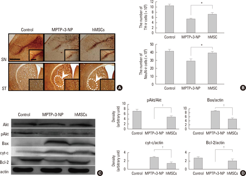
Fig. 2 Effects of cell therapy with human mesenchymal stem cells (hMSCs) on modulation of cell survival and death-signaling pathways in an animal model of multiple system atrophy-parkinsonism treated with MPTP and 3-NP. Immunohistochemical analysis showed that hMSC treatment significantly decreased the decline in the number of TH-ir and NeuN-ir cells in the substantia nigra (SN) and striatum (ST) of double-toxin treated animals (A). Stereological analysis revealed that the number of TH-ir and NeuN-ir cells was significantly higher in the hMSC-treated group than in the group treated with double toxin alone (B; n=5; *p<0.05). Scale bar: 100 µm. Western blot analysis, performed 4 weeks after first double-toxin injection, showed that the p-Akt expression was significantly decreased in double-toxin-treated mice compared with controls. However, hMSC administration in double-toxin-treated mice increased p-Akt expression. hMSC treatment significantly decreased Bax expression in double-toxin-treated mice, whereas hMSC treatment significantly increased the expression of Bcl-2 in these mice. In addition, hMSCs significantly decreased the expression of cytochrome c, which was elevated after double-toxin treatment. (C, D; n=3; †p<0.01). Ref. 16 with permission from Cognizant Communication Corporation.
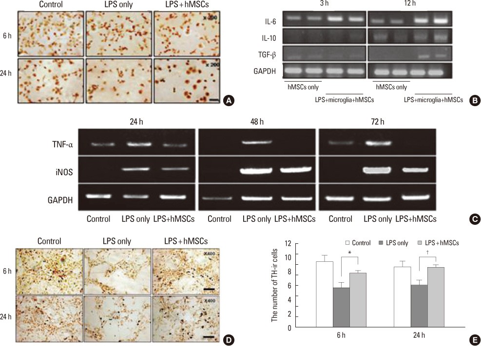
Fig. 3 Modulation of neuroinflammation by human mesenchymal stem cells (hMSCs) in co-culture systems. To identify soluble factors associated with modulation of microglial activation, we analyzed expression of IL-6, IL-10, and TGF-β in hMSCs co-cultured with LPS-stimulated microglia and hMSCs alone. The introduction of hMSCs significantly decreased the number of process-bearing activated microglia at 6 and 24 h following hMSC treatment (A). When hMSCs were co-cultured with LPS-stimulated microglia, IL-6 expression was significantly increased at 3 and 12 h, and IL-10 and TGF-β expressions were significantly increased at 12 h compared with hMSCs alone (B; n=5). Microglia from primary cultures were treated with LPS for 4 h and then co-cultured with vehicle or hMSCs in a Transwell. After 24, 48, and 72 h, culture supernatants and cells were collected for RT-PCR. LPS treatment significantly induced mRNA expression of TNF-α and iNOS compared with the control group, whereas co-culture with hMSCs showed significant reductions in TNF-α and iNOS mRNA expression (C) when compared with those treated with LPS alone at 24, 48, and 72 h. Co-cultures of microglia and mesencephalic neurons were treated with LPS for 4 h and then co-cultured with vehicle or hMSCs in a Transwell. LPS treatment resulted in a significant loss of TH-ir cells, whereas co-culture with hMSCs significantly decreased the loss of TH-ir cells (D). Stereological analysis revealed that the number of TH-ir cells was significantly higher in the hMSC-treated group than in the group treated with LPS alone (E; n=5, *p<0.05. †p<0.01). Scale bar: 100 µm. Ref. 17 with permission from Wiley.

Fig. 4 The protective effect of human mesenchymal stem cells (hMSCs) against lipopolysaccharide (LPS) and MPTP-induced damage to dopaminergic neurons in the substantia nigra (SN). The hMSCs treatment considerably reduced the loss of TH-ir cells and microglial activation induced by LPS stimulation in the SN (A). On stereological analysis, hMSC treatment significantly decreased the loss of TH-ir cells at 7 and 14 days following LPS stimulation (B; n=5, *p<0.05). The hMSC administration significantly downregulated the LPS-induced increase in the expression of TNF-α and iNOS mRNA at 3 days after LPS stimulation (C). Ref. 17 with permission from Wiley.
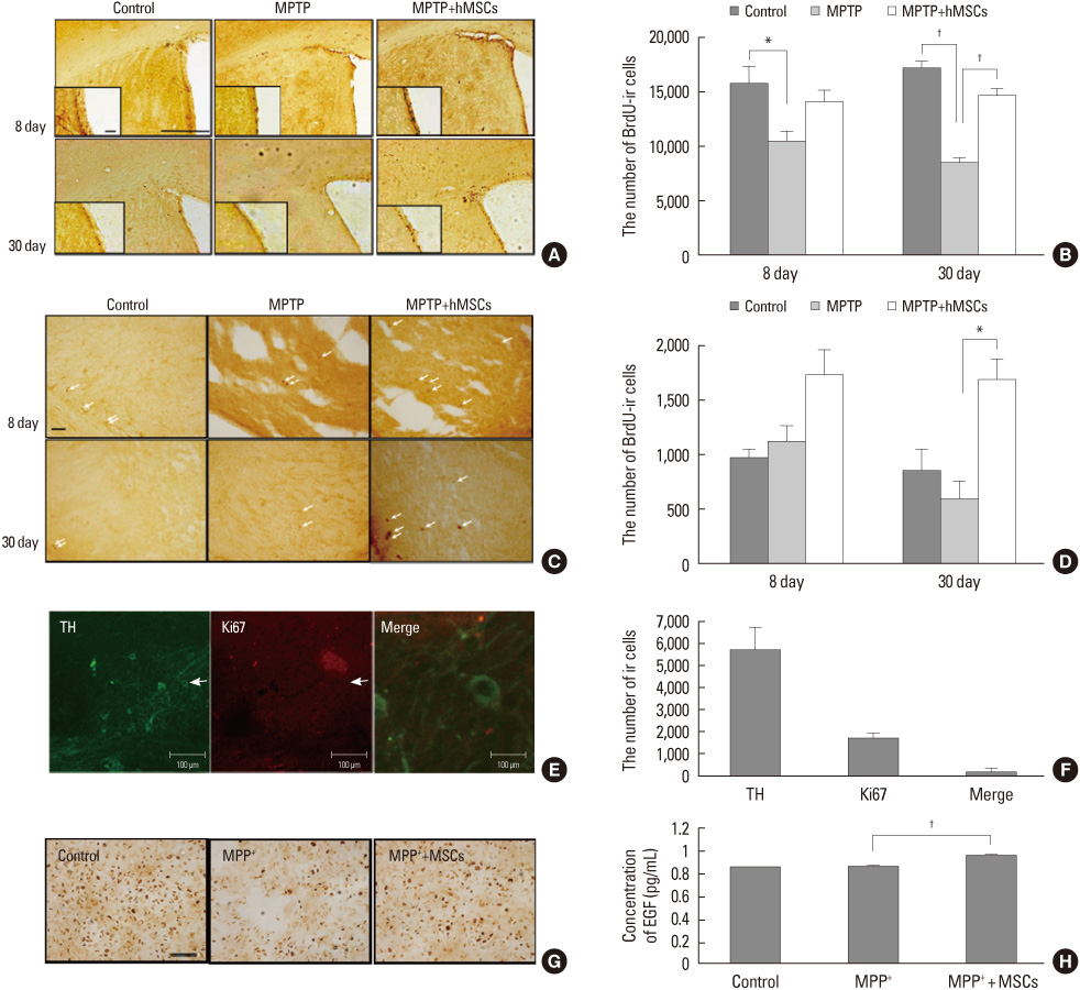
Fig. 5 Effect of human mesenchymal stem cells (hMSCs) on neurogenesis in Parkinsonian model. hMSC treatment prominently increased the number of BrdU-ir cells in the subventricular zone (SVZ) and substantia nigra (SN) of MPTP-treated PD animal models (A, C). Stereological analysis revealed that the number of BrdU-ir cells in the SVZ was significantly greater in the hMSCs-treated group compared with the MPTP-only-treated group at 30 days after MPTP injection (B, D; *p<0.05, †p<0.01). Brain tissue in the SN areas was double immunostained with Ki67 and TH at 22 days after last BrdU injection. Ki67 and TH-ir cells were not detected in the SN of MPTP-induced PD animal model; however, double-stained cells were notably observed in the SN of hMSCs-treated PD animals (E). On stereological analysis, the number of double-stained cells in the SN was 189±52, which roughly corresponded to 10% of the total number of Ki67-ir cells (F). To investigate the effect of hMSCs on the survival of NPCs obtained from the SVZ, the NPCs were cocultured with hMSCs using a Transwell after MPP+ treatment. The MPP+ administration induced marked loss of NPCs compared with the controls, whereas coculture with hMSCs resulted in a significant reduction in the loss of NPCs (G). The level of EGF in the medium of NPCs cotreated with hMSCs was significantly higher compared with that of MPP+-only treatment (H; n=3; †p<0.01). Scale bar: 100 µm. Ref. 24 with permission from Cognizant Communication Corporation.

Fig. 6 Neuroprotective effects of autologous mesenchymal stem cells in patients with multiple system atrophy. Changes in unified multiple system atrophy rating scale (UMSARS) scores from baseline through the follow-up period, according to treatment group. The graphs demonstrate a significant interaction effect between treatment group and time, indicating that the MSC-treated group had a much lesser increase in total UMSARS (A) and UMSARS II scores (B) compared with the placebo-treated group. Change in cerebral glucose metabolism and gray matter density from baseline (D0) to day 360 (D360) in the mesenchymal stem cell (MSC) and the placebo groups. Areas of decreased cerebral metabolism and gray matter density at D360 relative to D0 are illustrated in the MSC group (C-a and c) and placebo group (C-b and d). In both groups, cerebral glucose metabolism and gray matter density in the cerebellum were significantly decreased at 360 days compared with baseline. However, the placebo group showed more extensive areas of decreased cerebral glucose metabolism and gray matter density in the cerebellum and also in various cerebral cortex regions, particularly the frontal lobe, compared with the MSC group. The bars indicate the standard error. Ref. 25 with permission from Wiley.
Reference
-
1. Moore DJ, West AB, Dawson VL, Dawson TM. Molecular pathophysiology of Parkinson's disease. Annu Rev Neurosci. 2005. 28:57–87.
Article2. von Bohlen und Halbach O, Schober A, Krieglstein K. Genes, proteins, and neurotoxins involved in Parkinson's disease. Prog Neurobiol. 2004. 73:151–177.
Article3. Pittenger MF, Mackay AM, Beck SC, Jaiswal RK, Douglas R, Mosca JD, et al. Multilineage potential of adult human mesenchymal stem cells. Science. 1999. 284:143–147.
Article4. Woodbury D, Schwarz EJ, Prockop DJ, Black IB. Adult rat and human bone marrow stromal cells differentiate into neurons. J Neurosci Res. 2000. 61:364–370.
Article5. Minguell JJ, Erices A, Conget P. Mesenchymal stem cells. Exp Biol Med (Maywood). 2001. 226:507–520.
Article6. Bonuccelli U, Del Dotto P. New pharmacologic horizons in the treatment of Parkinson disease. Neurology. 2006. 67:S30–S38.
Article7. Barry FP, Murphy JM. Mesenchymal stem cells: clinical applications and biological characterization. Int J Biochem Cell Biol. 2004. 36:568–584.
Article8. Krampera M, Pasini A, Pizzolo G, Cosmi L, Romagnani S, Annunziato F. Regenerative and immunomodulatory potential of mesenchymal stem cells. Curr Opin Pharmacol. 2006. 6:435–441.
Article9. Karussis D, Kassis I, Kurkalli BG, Slavin S. Immunomodulation and neuroprotection with mesenchymal bone marrow stem cells (MSCs): a proposed treatment for multiple sclerosis and other neuroimmunological/neurodegenerative diseases. J Neurol Sci. 2008. 265:131–135.
Article10. Nauta AJ, Fibbe WE. Immunomodulatory properties of mesenchymal stromal cells. Blood. 2007. 110:3499–3506.
Article11. Blondheim NR, Levy YS, Ben-Zur T, Burshtein A, Cherlow T, Kan I, et al. Human mesenchymal stem cells express neural genes, suggesting a neural predisposition. Stem Cells Dev. 2006. 15:141–164.
Article12. Barzilay R, Kan I, Ben-Zur T, Bulvik S, Melamed E, Offen D. Induction of human mesenchymal stem cells into dopamine-producing cells with different differentiation protocols. Stem Cells Dev. 2008. 17:547–554.
Article13. Offen D, Barhum Y, Levy YS, Burshtein A, Panet H, Cherlow T, et al. Intrastriatal transplantation of mouse bone marrow-derived stem cells improves motor behavior in a mouse model of Parkinson's disease. J Neural Transm Suppl. 2007. 133–143.
Article14. Ye M, Wang XJ, Zhang YH, Lu GQ, Liang L, Xu JY, et al. Therapeutic effects of differentiated bone marrow stromal cell transplantation on rat models of Parkinson's disease. Parkinsonism Relat Disord. 2007. 13:44–49.
Article15. Park HJ, Lee PH, Bang OY, Lee G, Ahn YH. Mesenchymal stem cells therapy exerts neuroprotection in a progressive animal model of Parkinson's disease. J Neurochem. 2008. 107:141–151.
Article16. Park HJ, Bang G, Lee BR, Kim HO, Lee PH. Neuroprotective effect of human mesenchymal stem cells in an animal model of double toxin-induced multiple system atrophy parkinsonism. Cell Transplant. 2011. 20:827–835.
Article17. Kim YJ, Park HJ, Lee G, Bang OY, Ahn YH, Joe E, et al. Neuroprotective effects of human mesenchymal stem cells on dopaminergic neurons through anti-inflammatory action. Glia. 2009. 57:13–23.
Article18. Eriksson PS, Perfilieva E, Bjork-Eriksson T, Alborn AM, Nordborg C, Peterson DA, et al. Neurogenesis in the adult human hippocampus. Nat Med. 1998. 4:1313–1317.
Article19. Luskin MB. Restricted proliferation and migration of postnatally generated neurons derived from the forebrain subventricular zone. Neuron. 1993. 11:173–189.
Article20. Doetsch F, Hen R. Young and excitable: the function of new neurons in the adult mammalian brain. Curr Opin Neurobiol. 2005. 15:121–128.21. Borta A, Hoglinger GU. Dopamine and adult neurogenesis. J Neurochem. 2007. 100:587–595.22. Crews L, Mizuno H, Desplats P, Rockenstein E, Adame A, Patrick C, et al. Alpha-synuclein alters Notch-1 expression and neurogenesis in mouse embryonic stem cells and in the hippocampus of transgenic mice. J Neurosci. 2008. 28:4250–4260.
Article23. Winner B, Rockenstein E, Lie DC, Aigner R, Mante M, Bogdahn U, et al. Mutant alpha-synuclein exacerbates age-related decrease of neurogenesis. Neurobiol Aging. 2008. 29:913–925.
Article24. Park HJ, Shin JY, Lee BR, Kim HO, Lee PH. Mesenchymal stem cells augment neurogenesis in the subventricular zone and enhance differentiation of neural precursor cells into dopaminergic neurons in the substantia nigra of a Parkinsonian model. Cell Transplant. Forthcoming 2012.
Article25. Lee PH, Lee JE, Kim HS, Song SK, Lee HS, Nam HS, et al. A randomized trial of mesenchymal stem cells in multiple system atrophy. Ann Neurol. 2012. 72:32–40.
Article
- Full Text Links
-
- Actions
-
Cited
- CITED
-
- Close
- Share
-
- Similar articles
-
- Current Trends and Prospect of Cell Therapy using Hematopoietic Stem Cells
- Strategy for Maximizing Therapeutic Efficacy of Adult Stem Cells
- Adult Stem Cells: Beyond Regenerative Tool, More as a Bio-Marker in Obesity and Diabetes
- Stem Cells and Niemann Pick Disease
- Recent Trends and Strategies in Stem Cell Therapy for Alzheimer's Disease
