Differences in molar relationships and occlusal contact areas evaluated from the buccal and lingual aspects using 3-dimensional digital models
- Affiliations
-
- 1Department of Clinical Orthodontics, Graduate School of Clinical Dentistry, Ewha Womans University, Seoul, Korea. yschun@ewha.ac.kr
- KMID: 1435412
- DOI: http://doi.org/10.4041/kjod.2012.42.4.182
Abstract
OBJECTIVE
The aims of this study were to use a 3-dimensional (3D) system to compare molar relationship assessments performed from the buccal and lingual aspects, and to measure differences in occlusal contact areas between Class II and Class I molar relationships.
METHODS
Study casts (232 pairs from 232 subjects, yielding a total of 380 sides) were evaluated from both the buccal and lingual aspects, so that molar relationships could be classified according to the scheme devised by Liu and Melsen. Occlusal contact areas were quantified using 3D digital models, which were generated through surface scanning of the study casts.
RESULTS
A cusp-to-central fossa relationship was observed from the lingual aspect in the majority of cases classified from the buccal aspect as Class I (89.6%) or mild Class II (86.7%). However, severe Class II cases had lingual cusp-to-mesial triangular fossa or marginal ridge relationships. Mean occlusal contact areas were similar in the Class I and mild Class II groups, while the severe Class II group had significantly lower values than either of the other 2 groups (p < 0.05).
CONCLUSIONS
Buccal and lingual assessments of molar relationships were not always consistent. Occlusal contact areas were lowest for the Class II-severe group, which seems to have the worst molar relationships - especially as seen from the lingual aspect.
Keyword
MeSH Terms
Figure
-
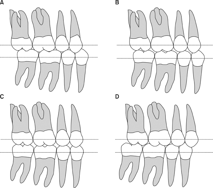
Figure 1 Guidelines used for classifying molar relationships from the buccal aspect. A, Class I; B, mild Class II (Class II-m); C, moderate Class II (Class II-m); D, severe Class II (Class II-s).
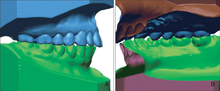
Figure 2 Sectioned images of 3-dimensional digital models showing the buccal and lingual surfaces of the first molars. These images were used to identify the position of the mesiopalatal cusp of the upper first molar with respect to the lower molar. A, Buccal aspect; B, lingual aspect.
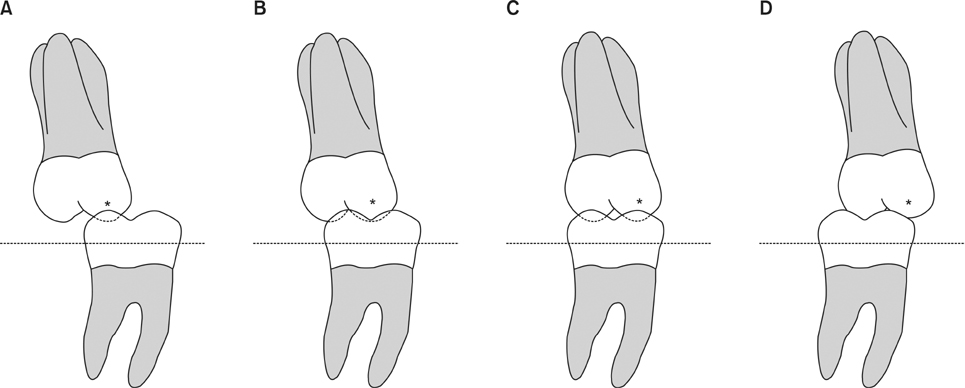
Figure 3 Guidelines used for classifying molar relationships from the lingual aspect. A, Lingual 0; B, lingual 1; C, lingual 2; D, lingual 3. *The mesiopalatal cusp of the upper first molar.
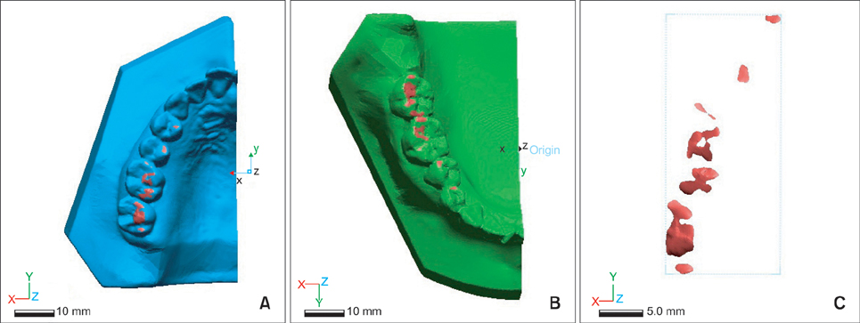
Figure 4 Three-dimensional digital models, produced by the Rapidform XOR3® program, showing the intersection areas (red) of the upper and lower scanned mesh. A, Upper right side; B, lower right side; C, occlusal contact area.
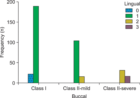
Figure 5 Distribution of samples (n) classified by evaluations from the buccal and lingual aspects. Lingual 0, distal triangular fossa; 1, central fossa; 2, mesial triangular fossa; 3, anterior aspect of the mesial triangular fossa.
Cited by 1 articles
-
Comparison of occlusal contact areas of class I and class II molar relationships at finishing using three-dimensional digital models
Hyejoon Lee, Minji Kim, Youn-Sic Chun
Korean J Orthod. 2015;45(3):113-120. doi: 10.4041/kjod.2015.45.3.113.
Reference
-
1. Liu D, Melsen B. Reappraisal of Class II molar relationships diagnosed from the lingual side. Clin Orthod Res. 2001. 4:97–104.
Article2. Casko JS, Vaden JL, Kokich VG, Damone J, James RD, Cangialosi TJ, et al. Objective grading system for dental casts and panoramic radiographs. American Board of Orthodontics. Am J Orthod Dentofacial Orthop. 1998. 114:589–599.3. Russell CW. An atlas of tooth form. 1969. 4th ed. Philadelphia: WB Saunders;12.4. Keating AP, Knox J, Bibb R, Zhurov AI. A comparison of plaster, digital and reconstructed study model accuracy. J Orthod. 2008. 35:191–201.
Article5. Cha BK, Choi JI, Jost-Brinkmann PG, Jeong YM. Applications of three-dimensionally scanned models in orthodontics. Int J Comput Dent. 2007. 10:41–52.6. Bishara SE, Hoppens BJ, Jakobsen JR, Kohout FJ. Changes in the molar relationship between the deciduous and permanent dentitions: a longitudinal study. Am J Orthod Dentofacial Orthop. 1988. 93:19–28.
Article7. Braun S, Kusnoto B, Evans CA. The effect of maxillary first molar derotation on arch length. Am J Orthod Dentofacial Orthop. 1997. 112:538–544.
Article8. Hansen GK, Caruso JM, West V, Andreiko CA, Farrage JR, Jeiroudi MT. The rotation of maxillary first molars, mandibular first molars, and maxillary first premolars in acceptable occlusions. Aust Orthod J. 1997. 14:242–246.9. Lamons FF, Holmes CW. The problem of the rotated maxillary first permanent molar. Am J Orthod. 1961. 47:246–272.
Article10. Moorrees CF, Gron AM, Lebret LM, Yen PK, Fröhlich FJ. Growth studies of the dentition: a review. Am J Orthod. 1969. 55:600–616.
Article11. Durbin DS, Sadowsky C. Changes in tooth contacts following orthodontic treatment. Am J Orthod Dentofacial Orthop. 1986. 90:375–382.
Article12. Gazit E, Lieberman MA. Occlusal contacts following orthodontic treatment. Measured by a photocclusion technique. Angle Orthod. 1985. 55:316–320.13. Luke DA, Lucas PW. Chewing efficiency in relation to occlusal and other variations in the natural human dentition. Br Dent J. 1985. 159:401–403.
Article14. van der Bilt A. Assessment of mastication with implications for oral rehabilitation: a review. J Oral Rehabil. 2011. 38:754–780.
Article15. Sierpinska T, Golebiewska M, Lapuc M. The effect of mastication on occlusal parameters in healthy volunteers. Adv Med Sci. 2008. 53:316–320.
Article16. Garcia Cartagena A, Gonzalez Sequeros O, Garrido Garcia VC. Analysis of two methods for occlusal contact registration with the T-Scan system. J Oral Rehabil. 1997. 24:426–432.
Article17. Garrido García VC, García Cartagena A, González Sequeros O. Evaluation of occlusal contacts in maximum intercuspation using the T-Scan system. J Oral Rehabil. 1997. 24:899–903.
Article18. Aras K, Hasanreisoğlu U, Shinogaya T. Masticatory performance, maximum occlusal force, and occlusal contact area in patients with bilaterally missing molars and distal extension removable partial dentures. Int J Prosthodont. 2009. 22:204–209.19. Choi YJ, Chung CJ, Kim KH. Changes in occlusal force and occlusal contact area after orthodontic treatment. Korean J Orthod. 2010. 40:176–183.
Article20. Sultana MH, Yamada K, Hanada K. Changes in occlusal force and occlusal contact area after active orthodontic treatment: a pilot study using pressure-sensitive sheets. J Oral Rehabil. 2002. 29:484–491.
Article21. Korn M. Aberrant eruption and its relationship to crowding: the need for early detection and management. Alpha Omegan. 1999. 92:19–27.
- Full Text Links
-
- Actions
-
Cited
- CITED
-
- Close
- Share
-
- Similar articles
-
- Comparison of occlusal contact areas of class I and class II molar relationships at finishing using three-dimensional digital models
- Photoelastic evaluation of mandibular posterior crossbite appliance
- Evaluation of mandibular cortical bone thickness for placement of temporary anchorage devices (TADs)
- Comparison of intraoral scanning and conventional impression techniques using 3-dimensional superimposition
- Analysis and evaluation of relative positions of mandibular third molar and mandibular canal impacts
