J Korean Bone Joint Tumor Soc.
2012 Dec;18(2):89-93. 10.5292/jkbjts.2012.18.2.89.
Muscle Infarction and Calcification of the Semitendinosus Tendon: A Case Report
- Affiliations
-
- 1Department of Orthopedic Surgery, Ilsan Paik Hospital, Inje University College of Medicine, Goyang, Korea. osd07@paik.ac.kr
- KMID: 1431676
- DOI: http://doi.org/10.5292/jkbjts.2012.18.2.89
Abstract
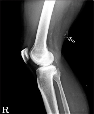
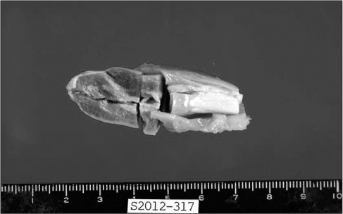
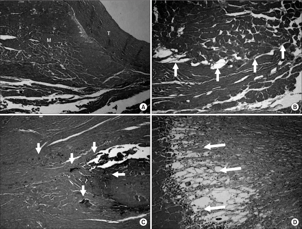
- The most common anatomic location of calcific tendinitis is the suprasupinatus muscle of the shoulder joint. However, it is known to develop in any joint including the hip, knee. Infarction of skeletal muscle in the distal areas of the limbs due to vascular occlusion is a well recognized systemic condition in patients who have diabetes. The author experienced mass-like lesion combined muscle infarction and calcification within pure semitendinosus tendon without diabetes in posterosuperior area of distal thigh in old age.
MeSH Terms
Figure
Reference
-
1. Uhthoff HK, Sarkar K, Maynard JA. Calcifying tendinitis: a new concept of its pathogenesis. Clin Orthop Relat Res. 1976. (118):164–168.2. Gärtner J, Simons B. Analysis of calcific deposits in calcifying tendinitis. Clin Orthop Relat Res. 1990. (254):111–120.3. Baudrillard JC, Lerais JM, Segal P, et al. Enthesopathy of the upper insertion of the musculus rectus femoris. A retrospective sign of tendon rupture in sports pathology. J Radiol. 1986. 67:185–191.4. Tennent TD, Goradia VK. Arthroscopic management of calcific tendinitis of the popliteus tendon. Arthroscopy. 2003. 19:E35.5. Aboulafia AJ, Monson DK, Kennon RE. Clinical and radiological aspects of idiopathic diabetic muscle infarction Rational approach to diagnosis and treatment. J Bone Joint Surg Br. 1999. 81:323–326.6. Damron TA, Levinsohn EM, McQuail TM, Cohen H, Stadnick M, Rooney M. Idiopathic necrosis of skeletal muscle in patients who have diabetes. Report of four cases and review of the literature. J Bone Joint Surg Am. 1998. 80:262–267.7. Lauro GR, Kissel JT, Simon SR. Idiopathic muscular infarction in a diabetic patient. Report of a case. J Bone Joint Surg Am. 1991. 73:301–304.8. Hinton A, Heinrich SD, Craver R. Idiopathic diabetic muscular infarction: the role of ultrasound, CT, MRI, and biopsy. Orthopedics. 1993. 16:623–625.9. Morcuende JA, Dobbs MB, Crawford H, Buckwalter JA. Diabetic muscle infarction. Iowa Orthop J. 2000. 20:65–74.
- Full Text Links
- Actions
-
Cited
- CITED
-
- Close
- Share
- Similar articles
-
- Chronic Tibialis Anterior Tendon Rupture Treated with Semitendinosus Autograft: A Report of Two Cases
- Reconstruction of Posterior Cruciate Ligament Using Semitendinosus Tendon (3 cases)
- Snapping Knee caused by the Semitendinous Tendon: A Case Report
- Isolated Semitendinosus Tendon Rupture in Non Athlete
- Free Semitendinosus Tendon Graft in Re-ruptured Achilles Tendon


