J Korean Soc Spine Surg.
2013 Jun;20(2):64-66. 10.4184/jkss.2013.20.2.64.
Acute Spinal Cord Infarction: Diffusion-Weighted MR Imaging: Case Report
- Affiliations
-
- 1Department of Orthopedic Surgery, School of Medicine, Wonkwang University, Iksan, Korea. niceo@hanmail.net
- 2Department of Neurology, School of Medicine, Wonwang University, Gunpo, Korea.
- 3Institute of Wonkwang Medical Science, Iksan, Korea.
- KMID: 1430377
- DOI: http://doi.org/10.4184/jkss.2013.20.2.64
Abstract
- STUDY DESIGN: A case report.
OBJECTIVE
We present a rare case of acute spinal cord infarction and usefulness of diffusion weighted MR imaging. SUMMARY OF LITERATURE REVIEW: T1-weighted and T2-weighted images are often normal in a patient with acute spinal cord infarction. MATERIAL AND METHODS: An 82-year-old presented with acute onset of paraplegia and urinary retention. His symptoms developed 6 days ago without any trauma. He had a history of vertebroplasty due to compression fracture of 12th thoracic vertebral body 6 years ago. There was no evidence of spinal cord compression on routine T1-and T2-weighted MRI.
RESULTS
In diffusion-weighted MRI, a high intensity signal intensity lesion in the spinal cord and conus medullaris was observed.
CONCLUSION
We report an example for the usefulness of diffusion-weighted image for early and accurate diagnosis of acute spinal cord infarction.
Keyword
MeSH Terms
Figure
-

Fig. 1. (A) T2-weighted sagittal image shows a strip like intramedullary high-signal intensity in the spinal cord. (B) and T1-weighted image shows no remarkable signal changes in the cord. (C) An axial T2-weighted image at the level of T12 show an symmetric intramedullary hyperintensy lesion. (D) An axial T1-weighted image at the level of T12 shows no abnormal signal changes.
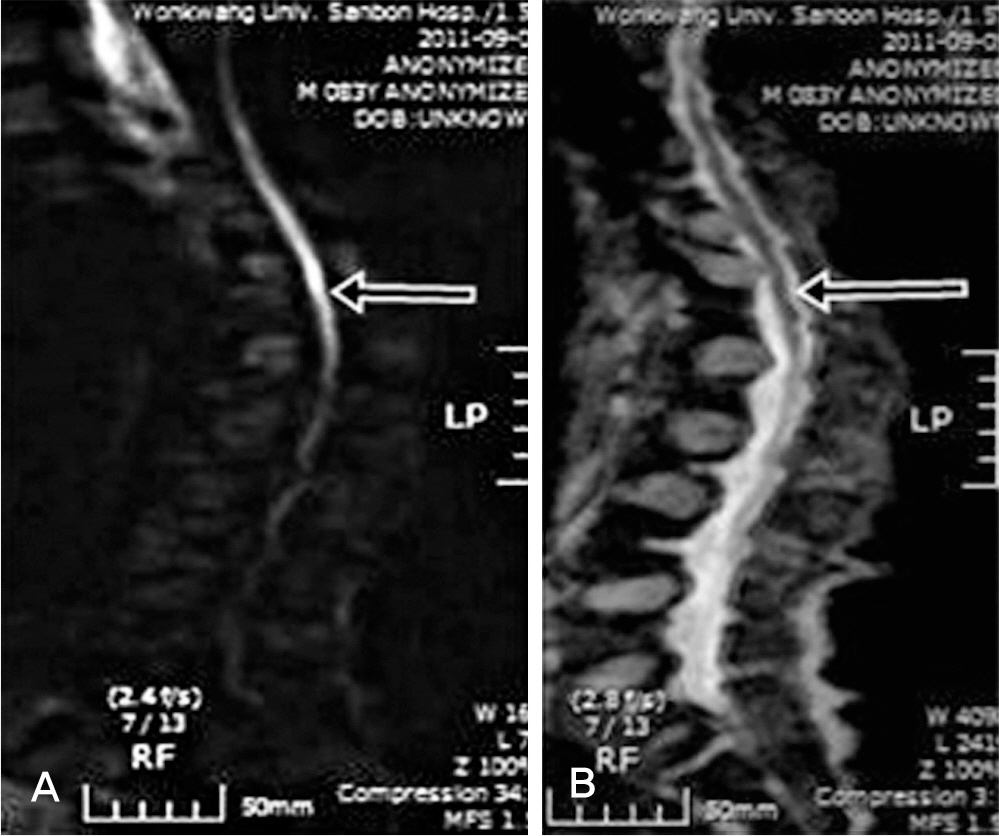
Fig. 2. (A) A sagittal diffusion-weighted image shows marked high signal intensity lesion at the corresponding area at the level of T12 (arrow) (B) The apparent diffusion coefficient (ADC) map image shows a low-signal intensity at the same area (arrow)
Reference
-
1. Loher TJ, Bassetti CL, Lö vblad KO, et al. Diffusion-weight-ed MRI in acute spinal cord ischaemia. Neuroradiology. 2003; 45:557–61.
Article2. Thurnher MM, Bammer R. Diffusion-weighted MR imaging (DWI) in spinal cord ischemia. Neuroradiology. 2006; 48:795–801.
Article3. Weidauer S, Nichtweiss M, Lanfermann H, Zanella FE. Spinal cord infarction: MR imaging and clinical features in 16 cases. Neuroradiology. 2002; 44:851–7.
Article4. Zhang J, Huan Y, Qian Y, Sun L, Ge Y. Multishot diffusion-weighted imaging features in spinal cord infarction. J Spinal Disord Tech. 2005; 18:277–82.5. de Figueiredo EH, Borgonovi AF, Doring TM. Basic con-cepts of MR imaging, diffusion MR imaging, and diffusion tensor imaging. Magn Reson Imaging Clin N Am. 2011; 19:1–22.
Article6. El-Koussy M, Lö vblad KO, Kiefer C, et al. Apparent diffusion coefficient mapping of infarcted tissue and the ischaemic penumbra in acute stroke. Neuroradiology. 2002; 44:812–8.
Article
- Full Text Links
-
- Actions
-
Cited
- CITED
-
- Close
- Share
-
- Similar articles
-
- Vertebral Artery Dissection Presenting with Acute Infarction in Cervical Spinal Cord and Cerebellum
- Diffusion-Weighted MR Imaging of Spinal Cord Infarction
- Delayed Post-Traumatic Spinal Cord Infarction with Quadriplegia: A Case Report
- The Role of Diffusion-Weighted MRI in Differentiation of Idiopathic Acute Transverse Myelitis and Acute Spinal Cord Infarction
- Usefulness of Diffusion-weighted MR Imaging in Acute Spinal Cord Infarction
