Restoration of Lumbar Lordosis After Posterior Lumbar Interbody Fusion with 4 Degree Cage in Degenerative Spinal Disease
- Affiliations
-
- 1Department of Orthopedic Surgery, School of Medicine, Inha University, Incheon, Korea. chokj@inha.ac.kr
- KMID: 1430375
- DOI: http://doi.org/10.4184/jkss.2013.20.2.51
Abstract
- STUDY DESIGN: Retrospective radiological evaluation.
OBJECTIVES
This purpose of this study is to determine how much lumbar lordosis and disc heights are restored after posterior lumbar interbody fusion (PLIF) with cage in degenerative spinal disease. SUMMARY OF LITERATURE REVIEW: Restoration of lumbar lordosis in lumbar spine surgery is crucial for clinical outcomes, but there are few studies about the relationship between restoration of lumbar lordosis and cage. MATERIAL AND METHOD: Eighty-one patients with degenerative spinal diseases underwent PLIF using metal cage with 4degrees lordotic angle. The mean age was 61 year-old (range 38-83 years). Cases with late complications including nonunion, subsidence of cage and instrument failure were excluded in this study. Lumbar lordosis, segmental lordosis, disc height, and sagittal alignment were analyzed on radiographs.
RESULTS
The fused level was one segment in 62 patients and two segments in 19 patients. All patients had the fusion from L3 to the sacrum. Preoperative lumbar lordosis was 34.2degrees, improved to 34.6degrees after surgery, and then changed to 32.2degrees at the final follow-up, demonstrating that the cage with 4degrees lordotic angle was not effective to restore lumbar lordosis. Segmental lordosis at the level of cage decreased at the final follow-up as compared to preoperative value at all segments. Disc height was improved at the final follow-up as compared to preoperative value.
CONCLUSION
Disc height was restored after PLIF using cage in the surgery for degenerative lumbar spine. However, lumbar lordosis and segmental lordosis were decreased at the final follow-up as compared to preoperative lordosis.
Figure
-
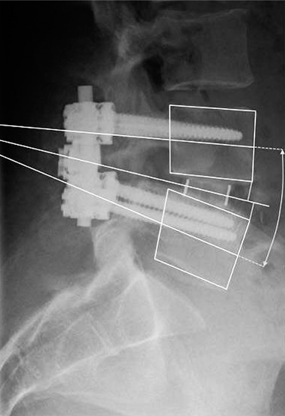
Fig. 1. Measurement method of disc height. The midpoint between the 2 posterior corners and the midpoints between the 2 anterior corners were computed. A bisectrix of the sagittal plane angle between the vertebra's midplane was computed and the perpendicular distances to the upper vertebra's lower anterior corner and the lower vertebra's upper anterior corner determined.
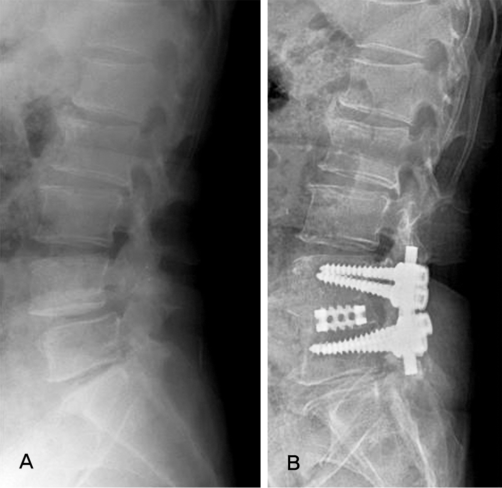
Fig 2. Loss of lumbar lordosis after posterior lumbar interbody fusion for the patient with spinal stenosis. (A) On preoperative x-ray lumbar lordosis was 32.7˚ and segmental lordosis at L45 was 7.4˚. (B) At the final followup, lumbar lordosis was 25.3˚ and segmental lordosis was 4.3˚ with significant loss of lordosis.
Cited by 1 articles
-
Lumbar Lordosis Restoration with an Eight Degree Cage in Posterior Lumbar Interbody Fusion for Lumbar Degenerative Disease
Young-Tae Kim, Kyu-Jung Cho, Ju-Yong Park, Jong-Hyuk Yang
J Korean Orthop Assoc. 2014;49(3):177-184. doi: 10.4055/jkoa.2014.49.3.177.
Reference
-
1. Cloward RB. The treatment of ruptured lumbar intervertebral discs by vertebral body fusion. I. Indications, operative technique, aftercare. J Neurosurg. 1953; 10:154–68.2. Collis JS. Total disc replacement. A modified posterior interbody fusion. Report of 750 cases. Clin Orthop Relat Res. 1985; 193:64–7.3. Henley EN Jr, David SM. Lumbar arthrodesis for the treatment of back pain. J bone Joint Surg Am. 1999; 81:716–30.4. Ma GW. Posterior lumbar interbody fusion with specialized instruments. Clin Orthop Relat Res. 1985; 193:57–63.
Article5. Arai Y, Takahashi M, Kurosawa H, Shitoto K. Comparative study of iliac bone graft and carbon cage with local bone graft in posterior lumbar interbody fusion. J Orthop Surg(Hong Kong). 2002; 10:1–7.
Article6. Bernhardt M, Bridwell KH. Segmental analysis of the sagittal plane alignment of the normal thoracic and lumbar spines and thoracolumbar junction. Spine (Phila Pa 1976). 1989; 14:717–21.
Article7. Lee CS, Oh WH, Chung SS, Lee SG, Lee JY. Analysis of the sagittal alignment of normal spines. J Korean Orthop Assoc. 1999; 34:949–54.
Article8. Liebensteiner MC, Jesacher G, Thaler M, Gstoettner M, Liebensteiner MV, Bach CM. Restoration and preservation of disc height and segmental lordosis with circumferential lumbar fusion: a retrospective analysis of cage versus bone graft. J Spinal Disord Tech. 2011; 24:44–9.9. Kim JH, Kim SS, Kim JH, Kim BJ. Comparison of mono-segment instrumented posterior lumbar interbody fusion with and without a metal cage in degenerative spine. J Korean Orthop Assoc. 2008; 43:143–51.
Article10. Song KJ, Lim YJ, Choi BW, Seo KB. Clinical efficacy of a stand-alone, threaded-titanium fusion cage for single-level degenerative lumbar spinal disorders. J Korean Orthop Assoc. 2008; 43:152–9.
Article11. Takahashi H, Suguro T, Yokoyama Y, Lida Y, Terashima F, Wada A. Effect of cage geometry on sagittal alignment after posterior lumbar interbody fusion for degenerative disc disease. J Orthop Surg(Hong Kong). 2010; 18:139–42.
Article12. Yoon YH, Cho KJ, Park SR, Moon KH, Lee TJ, Park HB. Posterior lumbar interbody fusion and unilateral posterolateral fusion with local bone and single cage- comparision to posterolateral lumbar fusion with autologous iliac bone. J Korean Orthop Assoc. 2009; 44:102–8.13. Hsieh PC, Koski TR, O'Shaughnessy BA, et al. Anterior lumbar interbody fusion in comparison with transforaminal lumbar interbody fusion: implications for the restoration of foraminal height, local disc angle, lumbar lordosis, and sagittal balance. J Neurosurg Spine. 2007; 7:379–86.
Article14. Cho KJ, Choi DH, Jung SR, Park SR. Local bone versus autogenous iliac bone graft for posterolateral fusion in the same patient. J Korean Soc Spine Surg. 2002; 9:211–5.15. Kroppenstedt S, Gulde M, Schonmayr R. Radiological comparison of instrumented posterior lumbar interbody fusion with one or two closed-box plasmapore coated tita-nium cages. Spine (Phila Pa 1976). 2008; 33:2083–8.
Article16. Groth A, Kuklo T, Klemme W, Polly D, Schroeder T. Comparison of sagittal contour and posterior disc height following interbody fusion; threaded cylindrical cages versus structural allograft versus vertical cages. J Spinal Disord. 2005; 18:332–6.
- Full Text Links
-
- Actions
-
Cited
- CITED
-
- Close
- Share
-
- Similar articles
-
- Lumbar Lordosis Restoration with an Eight Degree Cage in Posterior Lumbar Interbody Fusion for Lumbar Degenerative Disease
- Restoration of Segmental Lordosis and Related Factors in Interbody Fusion for Degenerative Lumbar Disease
- Minimally Invasive Lateral Lumbar Interbody Fusion: Indications, Outcomes and Complications
- Efficacy of Posterior Lumbar Interbody Fusion using PEEK Cage and Pedicle Screw Stabilization in Degenerative Lumbar Spinal Disorders: Minimum 3 Years Follow up Results
- Factors Affecting Segmental Lordotic Angle After Posterior Lumbar Interbody Fusion Using Metal Cage
