Clinical Comparison of the Predictive Value of the Simple Skull X-Ray and 3 Dimensional Computed Tomography for Skull Fractures of Children
- Affiliations
-
- 1Department of Neurosurgery, Ajou University School of Medicine, Suwon, Korea. ee802000@yahoo.co.kr
- KMID: 1426262
- DOI: http://doi.org/10.3340/jkns.2012.52.6.528
Abstract
OBJECTIVE
In the pediatric population the skull has not yet undergone ossification and it is assumed that the diagnostic rate of skull fractures by simple X-rays are lower than that of adults. It has been recently proposed that the diagnostic rates of skull fractures by 3-dimensional computer tomography (3D-CT) are higher than simple X-rays. The authors therefore attempted to compare the diagnostic rates of pediatric skull fractures by simple X-rays and 3D-CTs with respect to the type of fracture.
METHODS
One-hundred patients aged less than 12 years who visited the Emergency Center for cranial injury were subject to simple X-rays and 3D-CTs. The type and location of the fractures were compared and Kappa statistical analysis and the t-test were conducted.
RESULTS
Among the 100 pediatric patients, 65 were male and 35 were female. The mean age was 50+/-45 months. 63 patients had simple skull fractures and 22 had complex fractures, and the types of fractures were linear fractures in 74, diastatic fractures 15, depressed fractures in 10, penetrating fracture in 1, and greenstick fractures in 3 patients. Statistical difference was observed for the predictive value of simple skull fractures' diagnostic rate depending on the method for diagnosis. A significant difference of the Kappa value was noted in the diagnosis of depressed skull fractures and diastatic skull fractures.
CONCLUSION
In the majority of pediatric skull fractures, 3D-CT showed superior diagnosis rates compared to simple skull X-rays and therefore 3D-CT is recommended whenever skull fractures are suspected. This is especially true for depressed skull fractures and diastatic skull fractures.
Keyword
Figure
-
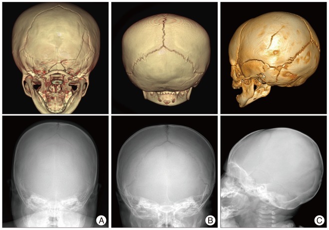
Fig. 1 Linear skull fracture in the simple X-ray and 3 dimensional computed tomography. Linear skull fractures on the occipital bone (A and B) and parietal bone (C) show not in the simple X-ray but clearly in the 3 dimensional computed tomography.
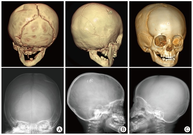
Fig. 2 Depressed skull fracture in the simple X-ray and 3 dimensional computed tomography. A : Depressed skull fracture on the parietal bone shows clearly both in the simple X-ray and 3 dimensional computed tomography. B and C : Depressed skull fracture on the outer table of the parietal bone shows not in the simple X-ray but clearly in the 3 dimensional computed tomography.
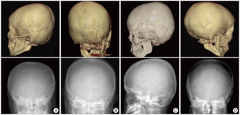
Fig. 3 Diastatic skull fractures in the simple X-ray and 3 dimensional computed tomography. A and B : Diastatic skull fractures on the lambdoid, temporo-parietal and occipito-mastoid sutures show clearly both in the simple X-ray and 3 dimensional computed tomography. C and D : Diastatic skull fractures on the coronal, lambdoid, and temporo-parietal sutures show not in the simple X-ray but clearly in the 3 dimensional computed tomography. Arrows indicate site of fracture in each pictures.
Reference
-
1. Brenner DJ, Hall EJ. Computed tomography--an increasing source of radiation exposure. N Engl J Med. 2007; 357:2277–2284. PMID: 18046031.
Article2. Choudhary AK, Jha B, Boal DK, Dias M. Occipital sutures and its variations : the value of 3D-CT and how to differentiate it from fractures using 3D-CT? Surg Radiol Anat. 2010; 32:807–816. PMID: 20174986.
Article3. Connor SE, Flis C. The contribution of high-resolution multiplanar reformats of the skull base to the detection of skull-base fractures. Clin Radiol. 2005; 60:878–885. PMID: 16039923.
Article4. Da Dalt L, Marchi AG, Laudizi L, Crichiutti G, Messi G, Pavanello L, et al. Predictors of intracranial injuries in children after blunt head trauma. Eur J Pediatr. 2006; 165:142–148. PMID: 16311740.
Article5. Di Scala C, Osberg JS, Gans BM, Chin LJ, Grant CC. Children with traumatic head injury : morbidity and postacute treatment. Arch Phys Med Rehabil. 1991; 72:662–666. PMID: 1830468.6. Gogos KA, Yakoumakis EN, Tsalafoutas IA, Makri TK. Radiation dose considerations in common paediatric X-ray examinations. Pediatr Radiol. 2003; 33:236–240. PMID: 12709751.
Article7. Jaffe D, Wesson D. Emergency management of blunt trauma in children. N Engl J Med. 1991; 324:1477–1482. PMID: 2023607.
Article8. Madeline LA, Elster AD. Suture closure in the human chondrocranium : CT assessment. Radiology. 1995; 196:747–756. PMID: 7644639.
Article9. Mann SS, Naidich TP, Towbin RB, Doundoulakis SH. Imaging of postnatal maturation of the skull base. Neuroimaging Clin N Am. 2000; 10:1–21. viiPMID: 10658152.10. Nelson EL, Melton LJ 3rd, Annegers JF, Laws ER, Offord KP. Incidence of skull fractures in Olmsted County, Minnesota. Neurosurgery. 1984; 15:318–324. PMID: 6332998.
Article11. Oh CK, Yoon SH. The significance of incomplete skull fracture in the birth injury. Med Hypotheses. 2010; 74:898–900. PMID: 20005051.
Article12. Quayle KS, Jaffe DM, Kuppermann N, Kaufman BA, Lee BC, Park TS, et al. Diagnostic testing for acute head injury in children : when are head computed tomography and skull radiographs indicated? Pediatrics. 1997; 99:E11. PMID: 9113968.13. Ringl H, Schernthaner RE, Schueller G, Balassy C, Kienzl D, Botosaneanu A, et al. The skull unfolded : a cranial CT visualization algorithm for fast and easy detection of skull fractures. Radiology. 2010; 255:553–562. PMID: 20332373.
Article14. Rutland-Brown W, Langlois JA, Thomas KE, Xi YL. Incidence of traumatic brain injury in the United States, 2003. J Head Trauma Rehabil. 2006; 21:544–548. PMID: 17122685.
Article15. Samii M, Tatagiba M. Skull base trauma : diagnosis and management. Neurol Res. 2002; 24:147–156. PMID: 11877898.16. Teasdale GM, Murray G, Anderson E, Mendelow AD, MacMillan R, Jennett B, et al. Risks of acute traumatic intracranial haematoma in children and adults : implications for managing head injuries. BMJ. 1990; 300:363–367. PMID: 2106986.
Article17. Viera AJ, Garrett JM. Understanding interobserver agreement : the kappa statistic. Fam Med. 2005; 37:360–363. PMID: 15883903.
- Full Text Links
-
- Actions
-
Cited
- CITED
-
- Close
- Share
-
- Similar articles
-
- Congenital depression of the neonatal skull unassociated with birth trauma
- Radiologic investigation of growing skull fracture
- Pneumocephalus in the Absence of Craniofacial Skull Base Fracture
- CT findings in patient with skull fractures
- Evaluation of the efficacy of simple skull examination in head trauma
