Non-Traumatic Myositis Ossificans in the Lumbosacral Paravertebral Muscle
- Affiliations
-
- 1Department of Neurosurgery, Ilsan Hospital, Dongguk University, Goyang, Korea. ktcho21@naver.com
- 2Department of Pathology, Ilsan Hospital, Dongguk University, Goyang, Korea.
- KMID: 1426201
- DOI: http://doi.org/10.3340/jkns.2013.53.5.305
Abstract
- Myositis ossificans (MO) is a benign condition of non-neoplastic heterotopic bone formation in the muscle or soft tissue. Trauma plays a role in the development of MO, thus, non-traumatic MO is very rare. Although MO may occur anywhere in the body, it is rarely seen in the lumbosacral paravertebral muscle (PVM). Herein, we report a case of non-traumatic MO in the lumbosacral PVM. A 42-year-old man with no history of trauma was referred to our hospital for pain in the low back, left buttock, and left thigh. On physical examination, a slightly tender, hard, and fixed mass was palpated in the left lumbosacral PVM. Computed tomography showed a calcified mass within the left lumbosacral PVM. Magnetic resonance imaging (MRI) showed heterogeneous high signal intensity in T1- and T2-weighted image, and no enhancement of the mass was found in the postcontrast T1-weighted MRI. The lack of typical imaging features required an open biopsy, and MO was confirmed. MO should be considered in the differential diagnosis when the imaging findings show a mass involving PVM. When it is difficult to distinguish MO from soft tissue or bone malignancy by radiology, it is necessary to perform a biopsy to confirm the diagnosis.
MeSH Terms
Figure
-
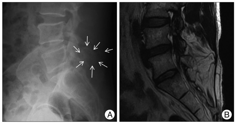
Fig. 1 Lateral X-ray and T2-weighted sagittal magnetic resonance imaging of the lumbosacral spine. A : Lateral lumbosacral spine X-ray shows calcific density near the spinous process of L5 and S1 (white arrows). B : T2-weighted sagittal magnetic resonance imaging shows heterogeneous high signal intensity at the paravetebral muscle from L5 to S2.

Fig. 2 Axial magnetic resonance imaging and computed tomography of the lumbosacral spine. A and B : T2- (A) and T1-weighted axial magnetic (B) resonance imaging shows a heterogeneous high signal intensity mass in the left paravetebral muscle. Neither edema nor low signal intensity in the surrounding tissue are found. C : T1-weghted postcontrast magnetic resonance imaging shows no enhancement of the mass. D : Computed tomography reveals an inhomogenously calcified mass, and the periphery of the mass is more calcified. The mass is in continuity with the left-side lamina, facet joint of the L5, and the sacrum.
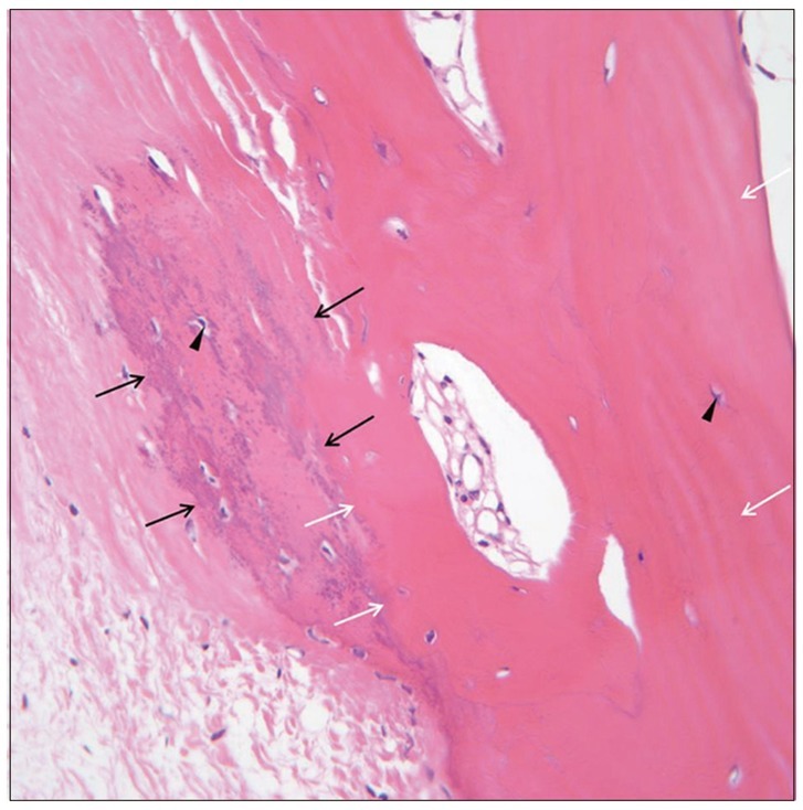
Fig. 3 Histopathologic study. Pathologic specimen shows osteocartilagenous tissue in the fibrous tissue and no malignant cell is found. A transition from immature woven bone (black arrows) to mature lamellar bone (white arrows) is present and normal osteocytes (arrow heads) are seen (Hematoxylin & Eosin stain, ×400).
Reference
-
1. Baysal T, Baysal O, Sarac K, Elmali N, Kutlu R, Ersoy Y. Cervical myositis ossificans traumatica : a rare location. Eur Radiol. 1999; 9:662–664. PMID: 10354880.2. Findlay I, Lakkireddi PR, Gangone R, Marsh G. A case of myositis ossificans in the upper cervical spine of a young child. Spine (Phila Pa 1976). 2010; 35:E1525–E1528. PMID: 21102285.
Article3. Hatano H, Morita T, Kobayashi H, Ito T, Segawa H. MR imaging findings of an unusual case of myositis ossificans presenting as a progressive mass with features of fluid-fluid level. J Orthop Sci. 2004; 9:399–403. PMID: 15278779.
Article4. Kim SW, Choi JH. Myositis ossificans in psoas muscle after lumbar spine fracture. Spine (Phila Pa 1976). 2009; 34:E367–E370. PMID: 19404167.
Article5. Kransdorf MJ, Meis JM, Jelinek JS. Myositis ossificans : MR appearance with radiologic-pathologic correlation. AJR Am J Roentgenol. 1991; 157:1243–1248. PMID: 1950874.
Article6. Mann SS, Som PM, Gumprecht JP. The difficulties of diagnosing myositis ossificans circumscripta in the paraspinal muscles of a human immunodeficiency virus-positive man : magnetic resonance imaging and temporal computed tomographic findings. Arch Otolaryngol Head Neck Surg. 2000; 126:785–788. PMID: 10864118.
Article7. Merchant R, Sainani NI, Lawande MA, Pungavkar SA, Patkar DP, Walawalkar A. Pre- and post-therapy MR imaging in fibrodysplasia ossificans progressiva. Pediatr Radiol. 2006; 36:1108–1111. PMID: 16932921.
Article8. Nishio J, Nabeshima K, Iwasaki H, Naito M. Non-traumatic myositis ossificans mimicking a malignant neoplasm in an 83-year-old woman : a case report. J Med Case Rep. 2010; 4:270. PMID: 20704714.9. Parikh J, Hyare H, Saifuddin A. The imaging features of post-traumatic myositis ossificans, with emphasis on MRI. Clin Radiol. 2002; 57:1058–1066. PMID: 12475528.
Article10. Saussez S, Blaivie C, Lemort M, Chantrain G. Non-traumatic myositis ossificans in the paraspinal muscles. Eur Arch Otorhinolaryngol. 2006; 263:331–335. PMID: 16133463.
Article11. Yazici M, Etensel B, Gürsoy MH, Aydoğdu A, Erkuş M. Nontraumatic myositis ossificans with an unusual location : case report. J Pediatr Surg. 2002; 37:1621–1622. PMID: 12407551.12. Zoccali C, Chichierchia G, Covello R. Regina Elena National Cancer Institute. An unusual case of lumbar paravertebral miositis ossificans mimicking muscular skeletal tumor. Musculoskelet Surg. 2011; 11. 12. [Epub ahead of print].
Article
- Full Text Links
-
- Actions
-
Cited
- CITED
-
- Close
- Share
-
- Similar articles
-
- Myositis Ossificans in Rectus Abdominis Muscle: Case Report
- Myositis ossificans progressiva
- A case of Non-Traumatic Myositis Ossificans in Quadriceps Femoris
- Myositis Ossificans of the Psoas Muscle After Compression Fracture of Lumbar Spine: CT and MR Imaging Findings
- Myositis Ossificans Progressiva: A Case Report
