J Korean Med Sci.
2012 Jun;27(6):614-618. 10.3346/jkms.2012.27.6.614.
Endothelial Dysfunction and Increased Carotid Intima-Media Thickness in the Patients with Slow Coronary Flow
- Affiliations
-
- 1The Heart Center of Chonnam National University Hospital, Gwangju, Korea. myungho@chollian.net
- 2Department of Nursing, Nambu University, Gwangju, Korea.
- KMID: 1421612
- DOI: http://doi.org/10.3346/jkms.2012.27.6.614
Abstract
- Flow mediated brachial dilatation (FMD) and carotid intima-media thickness (IMT) have been a surrogate for early atherosclerosis. Slow coronary flow in a normal coronary angiogram is not a rare condition, but its pathogenesis remains unclear. A total of 85 patients with angina were evaluated of their brachial artery FMD, carotid IMT and conventional coronary angiography. Coronary flow was quantified using the corrected thrombosis in myocardial infarction (TIMI) frame count method. Group I was a control with normal coronary angiography (n = 41, 56.1 +/- 8.0 yr) and group II was no significant coronary stenosis with slow flow (n = 44, 56.3 +/- 10.0 yr). Diabetes was rare but dyslipidemia and family history were frequent in group II. Heart rate was higher in group II than in group I. White blood cells, especially monocytes and homocysteine were higher in group II. The FMD was significantly lower in group II than in group I. Elevated heart rate, dyslipidemia and low FMD were independently related with slow coronary flow in regression analysis. Therefore, endothelial dysfunction may be an earlier vascular phenomenon than increased carotid IMT in the patients with slow coronary flow.
Keyword
MeSH Terms
-
Aged
Angina, Unstable/complications/physiopathology/ultrasonography
Brachial Artery/physiopathology
*Carotid Intima-Media Thickness
Coronary Angiography
Coronary Circulation/*physiology
Dyslipidemias/complications
Endothelium, Vascular/*physiopathology
Female
Heart Rate
Homocysteine/metabolism
Humans
Leukocyte Count
Male
Middle Aged
Monocytes/cytology
ROC Curve
Regression Analysis
Risk Factors
Homocysteine
Figure
-

Fig. 1 Differences of endothelial and vascular smooth muscular function. FMD, flow mediated dilatation; NMD, nitrogen mediated dilatation.
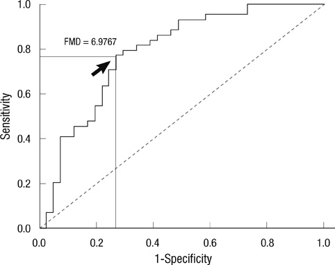
Fig. 2 Receiver operator characteristics curves of slow coronary flow. Arrow means cut off value of FMD (Area under the curve = 0.787, sensitivity = 0.773, specificity = 0.732).
Reference
-
1. Tambe AA, Demany MA, Zimmerman HA, Mascarenhas E. Angina pectoris and slow flow velocity of dye in coronary arteries: a new angiographic finding. Am Heart J. 1972. 84:66–71.2. Mangieri E, Macchiarelli G, Ciavolella M, Barillà F, Avella A, Martinotti A, Dell'ltalia LJ, Scibilia G, Motta P, Campa PP. Slow coronary flow: clinical and histopathological features in patients with otherwise normal epicardial coronary arteries. Cathet Cardiovasc Diagn. 1996. 37:375–381.3. Mosseri M, Yarom R, Gotsman MS, Hasin Y. Histologic evidence for small-vessel coronary artery disease in patients with angina pectoris and patent large coronary arteries. Circulation. 1986. 74:964–972.4. Singh S, Kothari SS, Bahl VK. Coronary slow flow phenomenon: an angiographic curiosity. Indian Heart J. 2004. 56:613–617.5. Simon A, Gariepy J, Chironi G, Megnien JL, Levenson J. Intima-media thickness: a new tool for diagnosis and treatment of cardiovascular risk. J Hypertens. 2002. 20:159–169.6. Roh EJ, Lim JW, Ko KO, Cheon EJ. A useful predictor of early atherosclerosis in obese children: serum high-sensitivity C-reactive protein. J Korean Med Sci. 2007. 22:192–197.7. Gibson CM, Cannon CP, Daley WL, Dodge JT Jr, Alexander B Jr, Marble SJ, McCabe CH, Raymond L, Fortin T, Poole WK, et al. TIMI frame count: a quantitative method of assessing coronary artery flow. Circulation. 1996. 93:879–888.8. Corretti MC, Anderson TJ, Benjamin EJ, CelermajerD , Charbonneau F, Creager MA, Deanfield J, Drexler H, Gerhard-Herman M, Herrington D, et al. Guidelines for the ultrasound assessment of endothelial-dependent flow-mediated vasodilation of the brachial artery: a report of the International Brachial Artery Reactivity Task Force. J Am Coll Cardiol. 2002. 39:257–265.9. Adams MR, Nakagomi A, Keech A, Robinson J, McCredie R, Bailey BP, Freedman SB, Celermajer DS. Carotid intima-media thickness is only weakly correlated with the extent and severity of coronary artery disease. Circulation. 1995. 92:2127–2134.10. Beltrame JF, Limaye SB, Wuttke RD, Horowitz JD. Coronary hemodynamic and metabolic studies of the coronary slow flow phenomenon. Am Heart J. 2003. 146:84–90.11. Gökçe M, Kaplan S, Tekelioğlu Y, Erdoğan T, Küçükosmanoğlu M. Platelet function disorder in patients with coronary slow flow. Clin Cardiol. 2005. 28:145–148.12. Cin VG, Pekdemir H, Camsar A, Ciçek D, Akkus MN, Parmaksýz T, Katýrcybaý T, Döven O. Diffuse intimal thickening of coronary arteries in slow coronary flow. Jpn Heart J. 2003. 44:907–919.13. Li JJ, Xu B, Li ZC, Qian J, Wei BQ. Is slow coronary flow associated with inflammation? Med Hypotheses. 2006. 66:504–508.14. Jin SM, Noh CI, Yang SW, Bae EJ, Shin CH, Chung HR, Kim YY, Yun YS. Endothelial dysfunction and microvascular complications in type 1 diabetes mellitus. J Korean Med Sci. 2008. 23:77–82.15. Sezgin N, Barutcu I, Sezgin AT, Gullu H, Turkmen M, Esen AM, Karakaya O. Plasma nitric oxide level and its role in slow coronary flow phenomenon. Int Heart J. 2005. 46:373–382.16. Rim SJ, Leong-Poi H, Lindner JR, Wei K, Fisher NG, Kaul S. Decrease in coronary blood flow reserve during hyperlipidemia is secondary to an increase in blood viscosity. Circulation. 2001. 104:2704–2709.17. Riza Erbay A, Turhan H, Yasar AS, Ayaz S, Sahin O, Senen K, Sasmaz H, Yetkin E. Elevated level of plasma homocysteine in patients with slow coronary flow. Int J Cardiol. 2005. 102:419–423.18. Fineschi M, Bravi A, Gori T. The "slow coronary flow" phenomenon: evidence of preserved coronary flow reserve despite increased resting microvascular resistances. Int J Cardiol. 2008. 127:358–361.19. Sezgin AT, Sigirci A, Barutcu I, Topal E, Sezgin N, Ozdemir R, Yetkin E, Tandogan I, Kosar F, Ermis N, et al. Vascular endothelial function in patients with slow coronary flow. Coron Artery Dis. 2003. 14:155–161.20. Wu WC, Sharma SC, Choudhary G, Coulter L, Coccio E, Eaton CB. Flow-mediated vasodilation predicts the presence and extent of coronary artery disease assessed by stress thallium imaging. J Nucl Cardiol. 2005. 12:538–544.21. Fineschi M, Gori T. Coronary slow flow: description of a new "cardiac Y" syndrome. Int J Cardiol. 2009. 137:308–310.22. Tanriverdi H, Evrengul H, Mergen H, Acar C, Seleci D, Kuru O, Tanriverdi S, Kaftan A. Early sign of atherosclerosis in slow coronary flow and relationship with angiotensin-converting enzyme I/D polymorphism. Heart Vessels. 2007. 22:1–8.23. Tanriverdi H, Evrengul H, Tanriverdi S, Kuru O, Seleci D, Enli Y, Kaftan A, Kilic M. Carotid intima-media thickness in coronary slow flow: relationship with plasma homocysteine levels. Coron Artery Dis. 2006. 17:331–337.
- Full Text Links
-
- Actions
-
Cited
- CITED
-
- Close
- Share
-
- Similar articles
-
- Carotid ultrasound in patients with coronary artery disease
- Slow Coronary Flow is Related to Increased Carotid Intima-Media Thickness but Not Pulse Wave Velocity
- Response: Increased Carotid Intima-Media Thickness Is Associated with Progression of Diabetic Nephropathy in Patients with Type 2 Diabetes
- Impact of Left Ventricular Ejection Fraction on Endothelial Function and Carotid Intima-Media Thickness in Patients with Coronary Artery Disease
- Do we need individual measurement of carotid intima and media thickness?
