Shear-Wave Elastography of Segmental Infarction of the Testis
- Affiliations
-
- 1Department of Radiology, Istanbul University, Cerrahpasa Medical Faculty, Istanbul 34300, Turkey. fatihkan@yahoo.com
- KMID: 1397516
- DOI: http://doi.org/10.3348/kjr.2012.13.6.820
Abstract
- Segmental testicular infarction (STI) is a rare cause of acute scrotum. The spectrum of findings on gray-scale and color Doppler ultrasonography differ depending on the time between the onset of testicular pain and the ultrasonography examination. We are not aware of the usefulness of shear-wave elastography for the diagnosis of STI. We report the shear-wave elastography features in a case of STI and discuss the role of this diagnostic modality in the differential diagnosis.
MeSH Terms
Figure
-
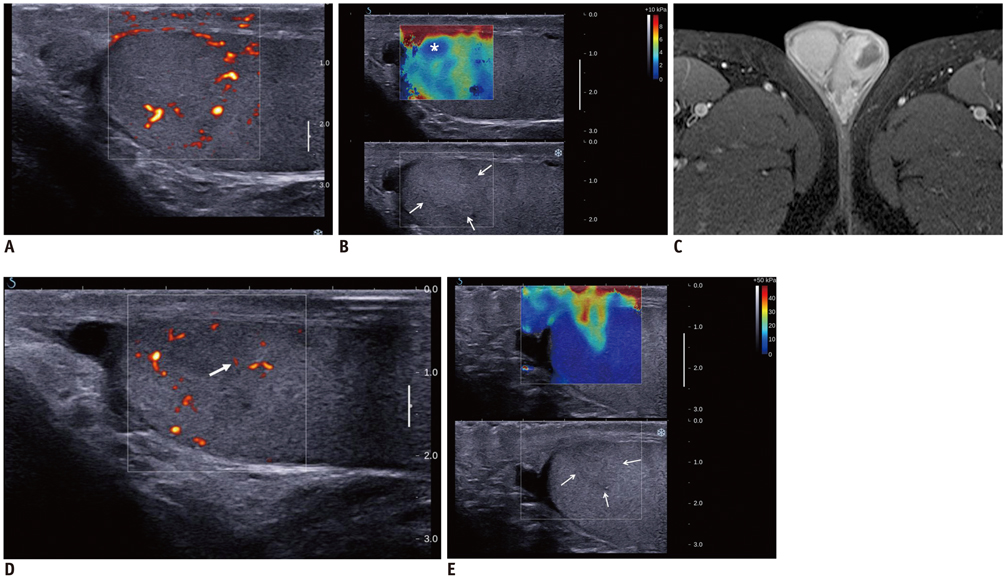
Fig. 1 Segmental testicular infarction in 35-year-old man. A. Power Doppler Ultrasonography (US) at initial presentation demonstrates absent flow in upper pole of left testis with perilesional hypervascularity. B. Gray-scale US of upper pole of left testis shows inconspicuous isohyperechoic almost round area (white arrows) with softer central part (asterisk) on shear-wave elastography (mean stiffness, 1.7 kPa). C. Gadolinium-enhanced magnetic resonance imaging reveals avascular area with perilesional hypervascularity (arrow). D, E. Follow-up US examinations were obtained 10 days later. Power Doppler US examination (D) demonstrates presence of intralesional vascularity (white arrow). Gray-scale and shear-wave elastography examinations (E) demonstrate that lesion is ill-defined but has wedge shape (white arrows) on gray-scale component of image. Elastography depicts more conspicuous triangular wedge shaped area corresponding to infarcted testicular lobule. Note that lesion is stiffer than normal testicular parenchyma.
Reference
-
1. Bilagi P, Sriprasad S, Clarke JL, Sellars ME, Muir GH, Sidhu PS. Clinical and ultrasound features of segmental testicular infarction: six-year experience from a single centre. Eur Radiol. 2007. 17:2810–2818.2. Secil M, Kocyigit A, Aslan G, Kefi A, Ozdemir I, Tuna B, et al. Segmental testicular infarction as a complication of varicocelectomy: sonographic findings. J Clin Ultrasound. 2006. 34:143–145.3. Fernández-Pérez GC, Tardáguila FM, Velasco M, Rivas C, Dos Santos J, Cambronero J, et al. Radiologic findings of segmental testicular infarction. AJR Am J Roentgenol. 2005. 184:1587–1593.4. Kim HK, Goske MJ, Bove KE, Minovich E. Segmental testicular infarction in a young man simulating a testicular tumor. Pediatr Radiol. 2009. 39:400–402.5. Athanasiou A, Tardivon A, Tanter M, Sigal-Zafrani B, Bercoff J, Deffieux T, et al. Breast lesions: quantitative elastography with supersonic shear imaging--preliminary results. Radiology. 2010. 256:297–303.6. Garra BS. Imaging and estimation of tissue elasticity by ultrasound. Ultrasound Q. 2007. 23:255–268.7. Goddi A, Sacchi A, Magistretti G, Almolla J, Salvadore M. Real-time tissue elastography for testicular lesion assessment. Eur Radiol. 2011. [Epub ahead of print].8. Bertolotto M, Derchi LE, Sidhu PS, Serafini G, Valentino M, Grenier N, et al. Acute segmental testicular infarction at contrast-enhanced ultrasound: early features and changes during follow-up. AJR Am J Roentgenol. 2011. 196:834–841.9. Sriprasad S, Kooiman GG, Muir GH, Sidhu PS. Acute segmental testicular infarction: differentiation from tumour using high frequency colour Doppler ultrasound. Br J Radiol. 2001. 74:965–967.10. Horstman WG, Melson GL, Middleton WD, Andriole GL. Testicular tumors: findings with color Doppler US. Radiology. 1992. 185:733–737.11. Sidhu PS, Sriprasad S, Bushby LH, Sellars ME, Muir GH. Impalpable testis cancer. BJU Int. 2004. 93:888.
- Full Text Links
-
- Actions
-
Cited
- CITED
-
- Close
- Share
-
- Similar articles
-
- Diagnostic Performance of Quantitative Shear Wave Ultrasound Elastography for Thyroid Cancer
- Future of breast elastography
- Shear wave elastography: a systematic review and meta-analysis
- Testicular stiffness in varicocele: evaluation with shear wave elastography
- Ultrasound elastography of the thyroid: principles and current status
