Radiation Dose Reduction of Chest CT with Iterative Reconstruction in Image Space - Part I: Studies on Image Quality Using Dual Source CT
- Affiliations
-
- 1Department of Radiology and Research Institute of Radiology, Asan Medical Center, University of Ulsan College of Medicine, Seoul 138-736, Korea. seojb@amc.seoul.kr
- 2Department of Radiology, Chungnam National University Hospital, Chungnam National University School of Medicine, Daejeon 301-721, Korea.
- 3Department of Radiology, Seoul St. Mary's Hospital, College of Medicine, The Catholic University of Korea, Seoul 137-701, Korea.
- KMID: 1397501
- DOI: http://doi.org/10.3348/kjr.2012.13.6.711
Abstract
OBJECTIVE
To determine whether the image quality (IQ) is improved with iterative reconstruction in image space (IRIS), and whether IRIS can be used for radiation reduction in chest CT.
MATERIALS AND METHODS
Standard dose chest CT (SDCT) in 50 patients and low dose chest CT (LDCT) in another 50 patients were performed, using a dual-source CT, with 120 kVp and same reference mAs (50 mAs for SDCT and 25 mAs for LDCT) employed to both tubes by modifying a dual-energy scan mode. Full-dose data were obtained by combining the data from both tubes and half-dose data were separated from a single tube. These were reconstructed by using a filtered back projection (FBP) and IRIS: full-dose FBP (F-FBP); full-dose IRIS (F-IRIS); half-dose FBP (H-FBP) and half-dose IRIS (H-IRIS). Objective noise was measured. The subjective IQ was evaluated by radiologists for the followings: noise, contrast and sharpness of mediastinum and lung.
RESULTS
Objective noise was significantly lower in H-IRIS than in F-FBP (p < 0.01). In both SDCT and LDCT, the IQ scores were highest in F-IRIS, followed by F-FBP, H-IRIS and H-FBP, except those for sharpness of mediastinum, which tended to be higher in FBP. When comparing CT images between the same dose and different reconstruction (F-IRIS/F-FBP and H-IRIS/H-FBP) algorithms, scores tended to be higher in IRIS than in FBP, being more distinct in half-dose images. However, despite the use of IRIS, the scores were lower in H-IRIS than in F-FBP.
CONCLUSION
IRIS generally helps improve the IQ, being more distinct at the reduced radiation. However, reduced radiation by half results in IQ decrease even when using IRIS in chest CT.
MeSH Terms
Figure
-

Fig. 1 Standard dose contrast enhanced-chest CT in 48-year-old woman (BMI: 23 kg/m2) with lung cancer in left upper lobe. A-C. Transverse CT images with (A) full radiation dose FBP in B70f kernel and 5 mm slice thickness, (B) full radiation dose IRIS in I70f kernel and 5 mm slice thickness, and (C) half radiation dose IRIS in I70f kernel and 5 mm slice thickness. Objective noise measured in saline bag was 66.1 HU, 37.5 HU and 53.8 HU, respectively (not shown). BMI = body mass index, HU = Hounsfield unit, FBP = filtered back projection, IRIS = iterative reconstruction in image space
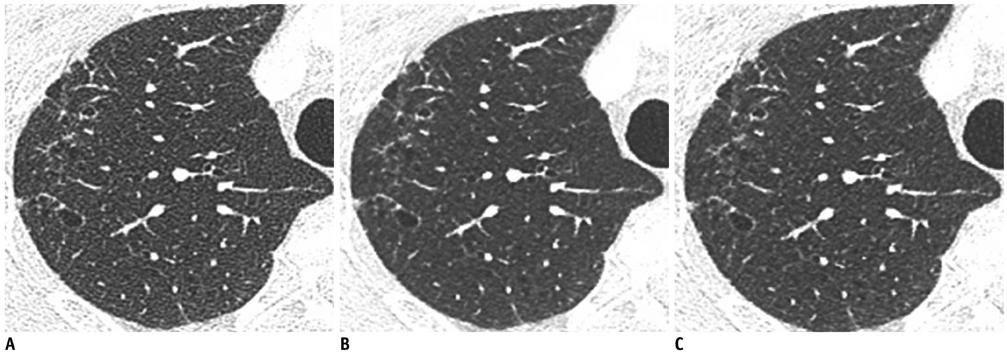
Fig. 2 Standard dose contrast enhanced-chest CT in 69-year-old man (BMI: 24 kg/m2) with mild emphysema. A-C. Transverse CT images with (A) full radiation dose FBP in B70f kernel and 1 mm slice thickness, (B) full radiation dose IRIS in I70f kernel and 1 mm slice thickness, and (C) half radiation dose with IRIS in I70f kernel and 1 mm slice thickness. Objective noise measured in saline bag was 59.9 HU, 35.7 HU and 47.6 HU, respectively (not shown). BMI = body mass index, HU = Hounsfield unit, FBP = filtered back projection, IRIS = iterative reconstruction in image space

Fig. 3 Low dose chest CT without enhancement in 37-year-old woman (BMI: 23 kg/m2) with small ground glass nodules. A-C. Transverse CT images with (A) full radiation dose FBP in B70f kernel and 5 mm slice thickness, (B) full radiation dose IRIS in I70f kernel and 5 mm slice thickness, and (C) half radiation dose IRIS in I70f kernel and 5 mm slice thickness. Objective noise measured in saline bag was 67.8 HU, 41.5 HU and 58.2 HU, respectively (not shown). BMI = body mass index, HU = Hounsfield unit, FBP = filtered back projection, IRIS = iterative reconstruction in image space
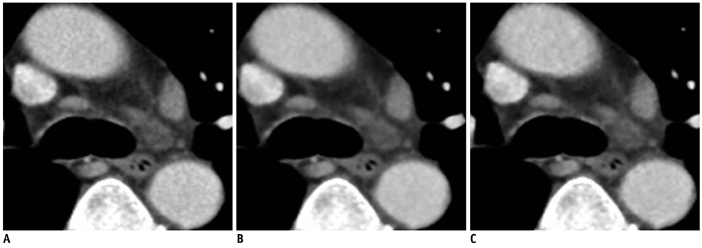
Fig. 4 Standard dose chest CT with enhancement in 75-year-old man (BMI: 21 kg/m2) with small lymph nodes in mediastinum. A-C. Transverse CT images with (A) full radiation dose FBP in B30f kernel and 5 mm slice thickness, (B) full radiation dose IRIS in I30f kernel and 5 mm slice thickness, and (C) half radiation dose IRIS in I30f kernel and 5 mm slice thickness. Objective noise measured in saline bag was 59.7 HU, 34.5 HU and 48.5 HU, respectively (not shown). Margins of mediastinal structures were slightly more blurred in (B) and (C) than in (A) due to excessive smoothing effect of IRIS image. BMI = body mass index, HU = Hounsfield unit, FBP = filtered back projection, IRIS = iterative reconstruction in image space
Cited by 1 articles
-
Effects of Dual-Energy CT with Non-Linear Blending on Abdominal CT Angiography
Sulan Li, Chaoqin Wang, Xiaochen Jiang, Ge Xu
Korean J Radiol. 2014;15(4):430-438. doi: 10.3348/kjr.2014.15.4.430.
Reference
-
1. Kalra MK, Maher MM, Toth TL, Schmidt B, Westerman BL, Morgan HT, et al. Techniques and applications of automatic tube current modulation for CT. Radiology. 2004. 233:649–657.2. Lell MM, May M, Deak P, Alibek S, Kuefner M, Kuettner A, et al. High-pitch spiral computed tomography: effect on image quality and radiation dose in pediatric chest computed tomography. Invest Radiol. 2011. 46:116–123.3. McNitt-Gray MF. AAPM/RSNA Physics Tutorial for Residents: topics in CT. Radiation dose in CT. Radiographics. 2002. 22:1541–1553.4. Mulkens TH, Bellinck P, Baeyaert M, Ghysen D, Van Dijck X, Mussen E, et al. Use of an automatic exposure control mechanism for dose optimization in multi-detector row CT examinations: clinical evaluation. Radiology. 2005. 237:213–223.5. Prasad SR, Wittram C, Shepard JA, McLoud T, Rhea J. Standard-dose and 50%-reduced-dose chest CT: comparing the effect on image quality. AJR Am J Roentgenol. 2002. 179:461–465.6. Rothenberg LN, Pentlow KS. Radiation dose in CT. Radiographics. 1992. 12:1225–1243.7. Schenzle JC, Sommer WH, Neumaier K, Michalski G, Lechel U, Nikolaou K, et al. Dual energy CT of the chest: how about the dose? Invest Radiol. 2010. 45:347–353.8. Wildberger JE, Mahnken AH, Schmitz-Rode T, Flohr T, Stargardt A, Haage P, et al. Individually adapted examination protocols for reduction of radiation exposure in chest CT. Invest Radiol. 2001. 36:604–611.9. Yu L, Li H, Fletcher JG, McCollough CH. Automatic selection of tube potential for radiation dose reduction in CT: a general strategy. Med Phys. 2010. 37:234–243.10. Dinkel HP, Sonnenschein M, Hoppe H, Vock P. Low-dose multislice CT of the thorax in follow-up of malignant lymphoma and extrapulmonary primary tumors. Eur Radiol. 2003. 13:1241–1249.11. Mayo JR, Hartman TE, Lee KS, Primack SL, Vedal S, Müller NL. CT of the chest: minimal tube current required for good image quality with the least radiation dose. AJR Am J Roentgenol. 1995. 164:603–607.12. Mayo JR, Kim KI, MacDonald SL, Johkoh T, Kavanagh P, Coxson HO, et al. Reduced radiation dose helical chest CT: effect on reader evaluation of structures and lung findings. Radiology. 2004. 232:749–756.13. Takahashi M, Maguire WM, Ashtari M, Khan A, Papp Z, Alberico R, et al. Low-dose spiral computed tomography of the thorax: comparison with the standard-dose technique. Invest Radiol. 1998. 33:68–73.14. Yamada T, Ono S, Tsuboi M, Saito H, Sato A, Matsuhashi T, et al. Low-dose CT of the thorax in cancer follow-up. Eur J Radiol. 2004. 51:169–174.15. Gregor J, Benson T. Computational analysis and improvement of SIRT. IEEE Trans Med Imaging. 2008. 27:918–924.16. Lasio GM, Whiting BR, Williamson JF. Statistical reconstruction for x-ray computed tomography using energy-integrating detectors. Phys Med Biol. 2007. 52:2247–2266.17. Xu J, Tsui BM. Electronic noise modeling in statistical iterative reconstruction. IEEE Trans Image Process. 2009. 18:1228–1238.18. AAPM. AAPM Report No. 96: the Measurement, Reporting, and Management of Radiation Dose in CT. 2008. College Park: American Association of Physicists in Medicine.19. Ravenel JG, Scalzetti EM, Huda W, Garrisi W. Radiation exposure and image quality in chest CT examinations. AJR Am J Roentgenol. 2001. 177:279–284.20. Pontana F, Duhamel A, Pagniez J, Flohr T, Faivre JB, Hachulla AL, et al. Chest computed tomography using iterative reconstruction vs filtered back projection (Part 2): image quality of low-dose CT examinations in 80 patients. Eur Radiol. 2011. 21:636–643.21. Pontana F, Pagniez J, Flohr T, Faivre JB, Duhamel A, Remy J, et al. Chest computed tomography using iterative reconstruction vs filtered back projection (Part 1): evaluation of image noise reduction in 32 patients. Eur Radiol. 2011. 21:627–635.22. Prakash P, Kalra MK, Digumarthy SR, Hsieh J, Pien H, Singh S, et al. Radiation dose reduction with chest computed tomography using adaptive statistical iterative reconstruction technique: initial experience. J Comput Assist Tomogr. 2010. 34:40–45.23. Hwang HJ, Seo JB, Lee JS, Song JW, Kim SS, Lee HJ, et al. Radiation dose reduction of chest CT with iterative reconstruction in image space - Part II: assessment of radiologists' preferences using dual source CT. Korean J Radiol. 2012. 13:720–727.24. Kyriakou Y, Kalender WA. Intensity distribution and impact of scatter for dual-source CT. Phys Med Biol. 2007. 52:6969–6989.
- Full Text Links
-
- Actions
-
Cited
- CITED
-
- Close
- Share
-
- Similar articles
-
- Radiation Dose Reduction of Chest CT with Iterative Reconstruction in Image Space - Part II: Assessment of Radiologists' Preferences Using Dual Source CT
- Quantitative Image Quality and Histogram-Based Evaluations of an Iterative Reconstruction Algorithm at Low-to-Ultralow Radiation Dose Levels: A Phantom Study in Chest CT
- Comparison of Radiation Dose and Image Quality between the 2nd Generation and 3rd Generation Dual-Source Single-Energy and Dual-Source Dual-Energy CT of the Abdomen
- Dose and Image Evaluations of Imaging for Radiotherapy
- Dosimetric Effects of Low Dose 4D CT Using a Commercial Iterative Reconstruction on Dose Calculation in Radiation Treatment Planning: A Phantom Study
