A Case of Cystoid Macular Edema Associated with Paclitaxel Chemotherapy
- Affiliations
-
- 1Department of Ophthalmology, Busan Paik Hospital, Inje University College of Medicine, Busan, Korea. maekbak@hanmail.net
- 2Department of Ophthalmology, Haeundae Paik Hospital, Inje University College of Medicine, Busan, Korea.
- KMID: 1397491
- DOI: http://doi.org/10.3341/kjo.2012.26.5.388
Abstract
- We encountered a patient with cystoid macular edema (CME) secondary to paclitaxel use. A 57-year-old man presented with gradual decreased bilateral vision. His chemotherapeutic regimen consisted of bevacizumab, paclitaxel (175 mg/m2 for 5 months), and carboplatin. Optical coherence tomography imaging revealed bilateral CME greater than 500 microm. However, one year later, visual acuity was improved, best-corrected Snellen visual acuity was 40 / 80 in each eye, and CME was spontaneously improved. Our study confirmed that macular edema associated with paclitaxel use shows spontaneous resolution and improvement of visual acuity after a change of chemotherapeutic regimen.
Keyword
MeSH Terms
-
Adenocarcinoma/drug therapy
Antineoplastic Agents, Phytogenic/*adverse effects
Antineoplastic Combined Chemotherapy Protocols/therapeutic use
Humans
Lung Neoplasms/drug therapy
Macular Edema/*chemically induced
Male
Middle Aged
Paclitaxel/*adverse effects
Remission, Spontaneous
Tomography, Optical Coherence
Visual Acuity
Figure
-
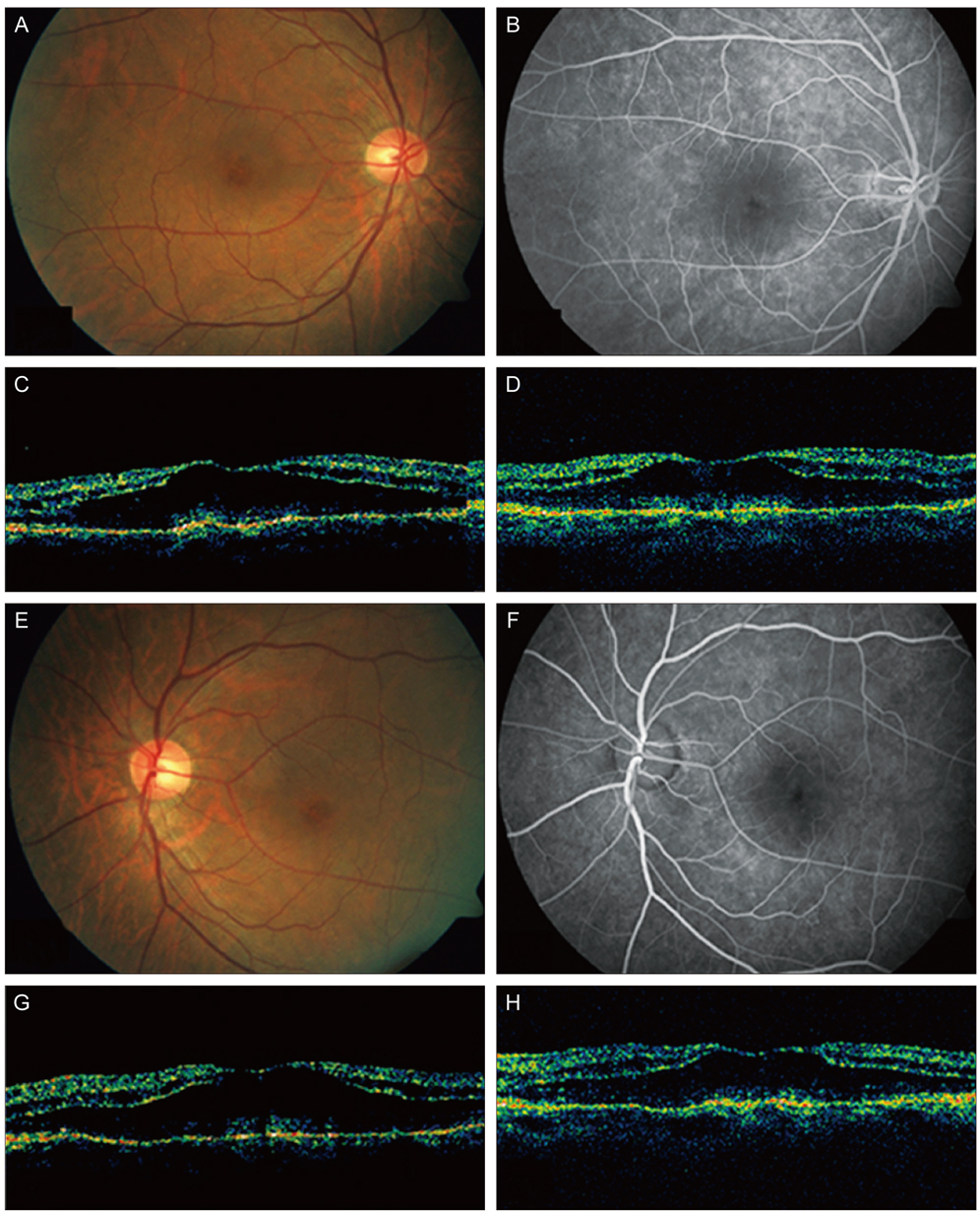
Fig. 1 (A-D) The right eye of the patient. (A) Before intravitreal bevacizumab plus triamcinolone injection, fundus showed cystic change of the macula. (B) Fluorescein angiograms (FA) showed hyperfluorescence in the macula in the late frame. (C) Optical coherence tomography (OCT) showed cystoid macular edema (CME). (D) Six weeks after the injection, CME was unchanged in OCT. (E-H) The left eye of the patient. (E) Before intravitreal bevacizumab plus triamcinolone injection, fundus showed cystic change of the macula. (F) FA showed hyperfluorescence in the macula in the late frame. (G) OCT showed CME. (H) Six weeks after the injection, CME was unchanged in OCT.

Fig. 2 The right (A) and left (B) eyes of the patient. One year later, optical coherence tomography showed cystoid macular edema improved. The central retinal thickness was decreased from 502 µm to 246 µm in the right eye and from 513 µm to 228 µm in the left eye.
Cited by 1 articles
-
Uveoretinal Adverse Effects Presented during Systemic Anticancer Chemotherapy: a 10-Year Single Center Experience
Ah Ran Cho, Young Hee Yoon, June-Gone Kim, Yoon Jeon Kim, Joo Yong Lee
J Korean Med Sci. 2018;33(7):. doi: 10.3346/jkms.2018.33.e55.
Reference
-
1. Tso MO. Pathology of cystoid macular edema. Ophthalmology. 1982. 89:902–915.2. Hofstra LS, de Vries EG, Willemse PH. Ophthalmic toxicity following paclitaxel infusion. Ann Oncol. 1997. 8:1053.3. Teitelbaum BA, Tresley DJ. Cystic maculopathy with normal capillary permeability secondary to docetaxel. Optom Vis Sci. 2003. 80:277–279.4. Telander DG, Sarraf D. Cystoid macular edema with docetaxel chemotherapy and the fluid retention syndrome. Semin Ophthalmol. 2007. 22:151–153.5. Joshi MM, Garretson BR. Paclitaxel maculopathy. Arch Ophthalmol. 2007. 125:709–710.
- Full Text Links
-
- Actions
-
Cited
- CITED
-
- Close
- Share
-
- Similar articles
-
- A Case of Paclitaxel-induced Maculopathy Treated with Methazolamide
- The Effect of Intravitreal Triamcinolone Acetonide Injection according to the Diabetic Macular Edema Type
- Comparison of Effects of IVTA and Photocoagulation, Depending on Types of Diabetic Macular Edema
- A Case of Secondary Macular Hole Formation after Phacoemulsification in a Vitrectomized Eye
- Prophylactic Intracameral Vancomycin Irrigation and Cystoid Macular Edema
