Morphologic Changes in Acute Central Serous Chorioretinopathy Using Spectral Domain Optical Coherence Tomography
- Affiliations
-
- 1Department of Ophthalmology, Konkuk University Medical Center, Konkuk University School of Medicine, Seoul, Korea. eyekim@kuh.ac.kr
- KMID: 1397485
- DOI: http://doi.org/10.3341/kjo.2012.26.5.347
Abstract
- PURPOSE
To investigate morphologic changes of acute central serous chorioretinopathy (CSC) using spectral domain optical coherence tomography (SD-OCT) and confocal scanning laser ophthalmoscopy.
METHODS
This retrospective study included 63 eyes of 63 patients with unilateral acute CSC. All patients underwent simultaneous SD-OCT and fluorescein angiography examination using Spectralis HRA+OCT.
RESULTS
The external limiting membrane could be seen on SD-OCT, although the junction between photoreceptor inner and outer segments (IS/OS) was not detected in all eyes with retinal detachment (RD). However, IS/OS became visible after resolution of serous RD in 51 eyes (81.0%). SD-OCT images at the leakage sites showed a bump of retinal pigment epithelium (RPE) in in 47 cases (68.1%) and pigment epithelial detachment (PED) in 22 of 69 leakage sites (31.9%). In 14 of 69 leakage sites (20.3%), highly reflective areas suggesting fibrinous exudate were observed in the subretinal space. In nine leakage sites (13.0%), sagging or dipping of the posterior retinal layer was seen. Abnormal RPE changes such as RPE bump and PED were observed in 12 of 22 fellow eyes (54.5%).
CONCLUSIONS
A variety of morphologic changes could be identified on SD-OCT, and those findings may contribute more information to our understanding of the pathophysiology of CSC.
Keyword
MeSH Terms
Figure
-

Fig. 1 Optical coherence tomography image of a 55-year-old woman with central serous chorioretinopathy. External limiting membrane (ELM) is clearly visible; however, the junction between photoreceptor inner and outer segments (IS/OS) is not detected in the detached neurosensory retina. The outer photoreceptor layer of the detached neurosensory retina above the clear subretinal space was irregularly thickened and granulated.
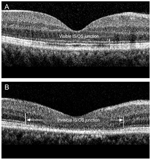
Fig. 2 Optical coherence tomography images of a 39-year-old man (A) and 46-year-old man (B) after resolution of serous detachment. (A) The junction of photoreceptor inner and outer segments (IS/OS) is intact and clearly visible. (B) IS/OS is disrupted and invisible.
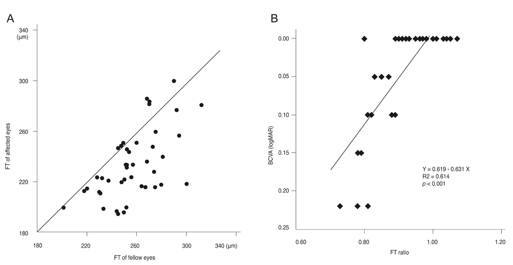
Fig. 3 (A) Distribution of foveal thickness (FT) of the affected eyes and the fellow eyes after resolution of serous detachment shows that most of the affected eyes have thinner FT than the fellow eyes. (B) FT ratio, the FT of the affected eye divided by that of the fellow eye, shows a significant positive correlation with best-corrected visual acuity (BCVA, logarithm of the minimum angle of resolution [logMAR]).

Fig. 4 Simultaneous imaging of fluorescein angiography and spectral domain optical coherence tomography (SD-OCT) in a 41-year-old man (A) and a 39-year-old man (B) with acute central serous chorioretinopathy. The horizontal linear scans of the SD-OCT image corresponding to the leakage sites show (A) pigment epithelial detachments (PED) and (B) retinal dipping above the retinal pigment epithelium (RPE) protrusion with fibrinous exudates in the subretinal space.
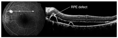
Fig. 5 Simultaneous imaging of fluorescein angiography (FA) and spectral domain optical coherence tomography (SD-OCT) in a 59-year-old woman with acute central serous chorioretinopathy. The minute defect in the retinal pigment epithelium (RPE) layer within the pigment epithelial detachment on SD-OCT corresponds exactly to a leakage point on FA.

Fig. 6 Simultaneous imaging of indocyanine green angiography (ICGA) and spectral domain optical coherence tomography in asymptomatic fellow eyes. An retinal pigment epithelium bump (A) and pigment epithelial detachment (B) are observed at the area of choroidal vascular hyperpermeability on ICGA.

Fig. 7 The spectral domain optical coherence tomographic findings of all the affected and fellow eyes in our study. RD = retinal detachment; ELM = external limiting membrane; IS/OS = inner and outer segments; RPE = retinal pigment epithelium; PED = pigment epithelial detachment.
Reference
-
1. Gass JD. Pathogenesis of disciform detachment of the neuroepithelium. Am J Ophthalmol. 1967. 63:Suppl. 1–139.2. Guyer DR, Yannuzzi LA, Slakter JS, et al. Digital indocyanine green videoangiography of central serous chorioretinopathy. Arch Ophthalmol. 1994. 112:1057–1062.3. Spaide RF, Campeas L, Haas A, et al. Central serous chorioretinopathy in younger and older adults. Ophthalmology. 1996. 103:2070–2079.4. Alam S, Zawadzki RJ, Choi S, et al. Clinical application of rapid serial fourier-domain optical coherence tomography for macular imaging. Ophthalmology. 2006. 113:1425–1431.5. Chen TC, Cense B, Pierce MC, et al. Spectral domain optical coherence tomography: ultra-high speed, ultra-high resolution ophthalmic imaging. Arch Ophthalmol. 2005. 123:1715–1720.6. Hangai M, Ojima Y, Gotoh N, et al. Three-dimensional imaging of macular holes with high-speed optical coherence tomography. Ophthalmology. 2007. 114:763–773.7. Ojima Y, Hangai M, Sasahara M, et al. Three-dimensional imaging of the foveal photoreceptor layer in central serous chorioretinopathy using high-speed optical coherence tomography. Ophthalmology. 2007. 114:2197–2207.8. Wojtkowski M, Bajraszewski T, Gorczynska I, et al. Ophthalmic imaging by spectral optical coherence tomography. Am J Ophthalmol. 2004. 138:412–419.9. Wojtkowski M, Srinivasan V, Fujimoto JG, et al. Three-dimensional retinal imaging with high-speed ultrahigh-resolution optical coherence tomography. Ophthalmology. 2005. 112:1734–1746.10. Wolf-Schnurrbusch UE, Enzmann V, Brinkmann CK, Wolf S. Morphologic changes in patients with geographic atrophy assessed with a novel spectral OCT-SLO combination. Invest Ophthalmol Vis Sci. 2008. 49:3095–3099.11. Iida T, Kishi S, Hagimura N, Shimizu K. Persistent and bilateral choroidal vascular abnormalities in central serous chorioretinopathy. Retina. 1999. 19:508–512.12. Piccolino FC, Borgia L. Central serous chorioretinopathy and indocyanine green angiography. Retina. 1994. 14:231–242.13. Prunte C, Flammer J. Choroidal capillary and venous congestion in central serous chorioretinopathy. Am J Ophthalmol. 1996. 121:26–34.14. Scheider A, Nasemann JE, Lund OE. Fluorescein and indocyanine green angiographies of central serous choroidopathy by scanning laser ophthalmoscopy. Am J Ophthalmol. 1993. 115:50–56.15. Hussain N, Baskar A, Ram LM, Das T. Optical coherence tomographic pattern of fluorescein angiographic leakage site in acute central serous chorioretinopathy. Clin Experiment Ophthalmol. 2006. 34:137–140.16. Iida T, Hagimura N, Sato T, Kishi S. Evaluation of central serous chorioretinopathy with optical coherence tomography. Am J Ophthalmol. 2000. 129:16–20.17. Kamppeter B, Jonas JB. Central serous chorioretinopathy imaged by optical coherence tomography. Arch Ophthalmol. 2003. 121:742–743.18. Mitarai K, Gomi F, Tano Y. Three-dimensional optical coherence tomographic findings in central serous chorioretinopathy. Graefes Arch Clin Exp Ophthalmol. 2006. 244:1415–1420.19. Fujimoto H, Gomi F, Wakabayashi T, et al. Morphologic changes in acute central serous chorioretinopathy evaluated by fourier-domain optical coherence tomography. Ophthalmology. 2008. 115:1494–1500. 1500.e1–1500.e2.20. Matsumoto H, Kishi S, Otani T, Sato T. Elongation of photoreceptor outer segment in central serous chorioretinopathy. Am J Ophthalmol. 2008. 145:162–168.21. Jalkh AE, Jabbour N, Avila MP, et al. Retinal pigment epithelium decompensation. I. Clinical features and natural course. Ophthalmology. 1984. 91:1544–1548.22. Yannuzzi LA, Shakin JL, Fisher YL, Altomonte MA. Peripheral retinal detachments and retinal pigment epithelial atrophic tracts secondary to central serous pigment epitheliopathy. Ophthalmology. 1984. 91:1554–1572.23. Matsumoto H, Sato T, Kishi S. Outer nuclear layer thickness at the fovea determines visual outcomes in resolved central serous chorioretinopathy. Am J Ophthalmol. 2009. 148:105–110.e1.24. Montero JA, Ruiz-Moreno JM. Optical coherence tomography characterisation of idiopathic central serous chorioretinopathy. Br J Ophthalmol. 2005. 89:562–564.25. Shinojima A, Hirose T, Mori R, et al. Morphologic findings in acute central serous chorioretinopathy using spectral domain-optical coherence tomography with simultaneous angiography. Retina. 2010. 30:193–202.26. Perkins SL, Kim JE, Pollack JS, Merrill PT. Clinical characteristics of central serous chorioretinopathy in women. Ophthalmology. 2002. 109:262–266.27. Eandi CM, Ober M, Iranmanesh R, et al. Acute central serous chorioretinopathy and fundus autofluorescence. Retina. 2005. 25:989–993.
- Full Text Links
-
- Actions
-
Cited
- CITED
-
- Close
- Share
-
- Similar articles
-
- Spectral Domain OCT Findings of Asymptomatic Fellow Eyes in Central Serous Chorioretinopathy
- Effect of Serous Retinal Detachment on the Measurement of Axial Length in Central Serous Chorioretinopathy
- The Use fulness of OCT[Optical Coherence Tomography]for the Diagnosis of Central Serous Choriore tinopathy
- Central Serous Chorioretinopathy in a Patient with Retinal Macrovessel
- Subfoveal Choroidal Thickness in Fellow Eyes of Patients with Central Serous Chorioretinopathy
