Partially Cystic Thyroid Nodules: Ultrasound Findings of Malignancy
- Affiliations
-
- 1Department of Radiology, Kangbuk Samsung Hospital, Sungkyunkwan University School of Medicine, Seoul 110-746, Korea. radiokwag@hanmail.net
- KMID: 1392929
- DOI: http://doi.org/10.3348/kjr.2012.13.5.530
Abstract
OBJECTIVE
To seek for the ultrasound (US) findings of partially cystic thyroid nodules that are associated with malignancy.
MATERIALS AND METHODS
We reviewed the US characteristics of 22 surgically confirmed partially cystic papillary carcinomas, and compared them with those of 80 benign partially cystic nodules. The review cases were selected in a random order from a total of 1029 partially cystic nodules that were diagnosed with an US-guided fine needle aspiration biopsy over a period of 8 years (June 2003 to October 2010) at our institution.
RESULTS
In partially cystic thyroid nodules, a taller-than-wide shape (100%, p < 0.001) and spiculated or microlobulated margin (58.3%, p = 0.003) were significantly associated with malignancy. In terms of internal solid portion of the nodule, eccentric configuration (68.0%, p < 0.001), non-smooth margin (81.3%, p < 0.001), hypoechogenecity (30.0%, p < 0.042), and microcalcification (89.5%, p < 0.001) were more frequently demonstrated in malignant nodules than benign ones.
CONCLUSION
In partially cystic thyroid nodules, understanding the characteristics of US findings is important to make a precise diagnosis of malignant nodules.
Keyword
MeSH Terms
-
Adolescent
Adult
Aged
Biopsy, Fine-Needle
Carcinoma, Papillary/pathology/surgery/*ultrasonography
Case-Control Studies
Chi-Square Distribution
Cysts/pathology/surgery/*ultrasonography
Female
Humans
Male
Middle Aged
Statistics, Nonparametric
Thyroid Neoplasms/pathology/surgery/*ultrasonography
Thyroid Nodule/pathology/surgery/*ultrasonography
Figure
-
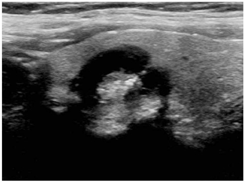
Fig. 1 Longitudinal ultrasound image of papillary thyroid carcinoma in 53-year-old woman shows predominantly cystic nodule. Entire nodule has smooth margin. Internal protruding eccentric solid position contains multiple punctuate round echogenic foci suggesting microcalcifications.
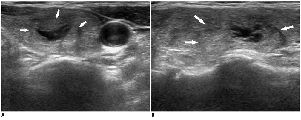
Fig. 2 Example of non-eccentric configuration of internal solid portion. Transverse (A) and longitudinal (B) US images show partially cystic nodule with isoechoic internal solid portion in 57-year-old woman. Entire nodule has ill-defined margin (arrows). Nodular hyperplasia was diagnosed by US-guided fine needle aspiration. US = ultrasound
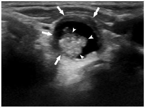
Fig. 3 Transverse ultrasound image of predominantly cystic nodule in 69-year-old woman. Eccentric solid position protruded internally and contained multiple microcalcifications. Note difference between smooth margin of entire nodule (arrows) and non-smooth margin of internal solid portion (arrowheads). Papillary thyroid carcinoma was diagnosed by fine needle aspiration and surgery.
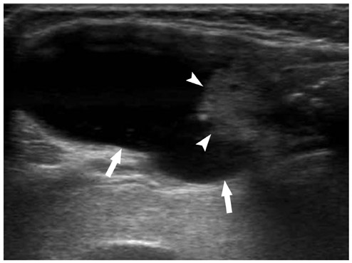
Fig. 4 Longitudinal ultrasound image of predominantly cystic nodule in 66-year-old woman shows eccentric configuration. Note difference between smooth margin of entire nodule (arrows) and non-smooth margin of internal solid portion (arrowheads). This lesion was surgically confirmed as papillary thyroid carcinoma despite substantial cystic portion.
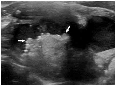
Fig. 5 Transverse ultrasound image of predominantly cystic nodule in 63-year-old woman shows eccentric configuration of internal solid portion with multiple microcalcifications. Note non-smooth margin of internal solid portion (arrows). This lesion was surgically confirmed as papillary thyroid carcinoma despite substantial cystic portion.
Cited by 1 articles
-
Complementary Role of Elastography Using Carotid Artery Pulsation in the Ultrasonographic Assessment of Thyroid Nodules: A Prospective Study
Soo Yeon Hahn, Jung Hee Shin, Eun Young Ko, Jung Min Bae, Ji Soo Choi, Ko Woon Park
Korean J Radiol. 2018;19(5):992-999. doi: 10.3348/kjr.2018.19.5.992.
Reference
-
1. McHenry CR, Slusarczyk SJ, Khiyami A. Recommendations for management of cystic thyroid disease. Surgery. 1999. 126:1167–1171. discussion 1171-1172.2. Khoo ML, Asa SL, Witterick IJ, Freeman JL. Thyroid calcification and its association with thyroid carcinoma. Head Neck. 2002. 24:651–655.3. do Rosário PW, Fagundes TA, Maia FF. Ultrasonographic features of papillary thyroid carcinoma. J Ultrasound Med. 2004. 23:572.4. Peccin S, de Castsro JA, Furlanetto TW, Furtado AP, Brasil BA, Czepielewski MA. Ultrasonography: is it useful in the diagnosis of cancer in thyroid nodules? J Endocrinol Invest. 2002. 25:39–43.5. Kim DW, Lee EJ, In HS, Kim SJ. Sonographic differentiation of partially cystic thyroid nodules: a prospective study. AJNR Am J Neuroradiol. 2010. 31:1961–1966.6. Lee MJ, Kim EK, Kwak JY, Kim MJ. Partially cystic thyroid nodules on ultrasound: probability of malignancy and sonographic differentiation. Thyroid. 2009. 19:341–346.7. Moon WJ, Baek JH, Jung SL, Kim DW, Kim EK, Kim JY, et al. Ultrasonography and the ultrasound-based management of thyroid nodules: consensus statement and recommendations. Korean J Radiol. 2011. 12:1–14.8. Kim SH, Kim BS, Jung SL, Lee JW, Yang PS, Kang BJ, et al. Ultrasonographic findings of medullary thyroid carcinoma: a comparison with papillary thyroid carcinoma. Korean J Radiol. 2009. 10:101–105.9. Gharib H, Papini E, Paschke R, Duick DS, Valcavi R, Hegedüs L, et al. American Association of Clinical Endocrinologists, Associazione Medici Endocrinologi, and EuropeanThyroid Association Medical Guidelines for Clinical Practice for the Diagnosis and Management of Thyroid Nodules. Endocr Pract. 2010. 16:Suppl 1. 1–43.10. Kim EK, Park CS, Chung WY, Oh KK, Kim DI, Lee JT, et al. New sonographic criteria for recommending fine-needle aspiration biopsy of nonpalpable solid nodules of the thyroid. AJR Am J Roentgenol. 2002. 178:687–691.11. Moon WJ, Jung SL, Lee JH, Na DG, Baek JH, Lee YH, et al. Benign and malignant thyroid nodules: US differentiation--multicenter retrospective study. Radiology. 2008. 247:762–770.12. Lee YH, Kim DW, In HS, Park JS, Kim SH, Eom JW, et al. Differentiation between benign and malignant solid thyroid nodules using an US classification system. Korean J Radiol. 2011. 12:559–567.13. Hiromura T, Nojima T, Morita Y, Choji K, Nakada K, Tsukamoto E, et al. Cystic papillary carcinoma of the thyroid--sonographic-pathologic correlation. Nihon Igaku Hoshasen Gakkai Zasshi. 1990. 50:40–47.14. Oertel YC, Oertel JE. Wartofsky L, Nostrand D, editors. Papillary Carcinoma: Cytology and Pathology. Thyroid Cancer. 2006. Totowa: Humana Press;263–270.
- Full Text Links
-
- Actions
-
Cited
- CITED
-
- Close
- Share
-
- Similar articles
-
- Thyroid nodules with minimal cystic changes have a low risk of malignancy
- US Diagnosis for Thyroid Nodules with an Indeterminate Cytology
- Malignancy risk of thyroid nodules with minimal cystic changes: a multicenter retrospective study
- Malignancy Risk Stratification of Thyroid Nodules with Macrocalcification and Rim Calcification Based on Ultrasound Patterns
- Ultrasonographic Findings of Thyroid Nodules: Differentiation between Malignant and Benign Nodules
