Korean J Ophthalmol.
2012 Aug;26(4):271-276. 10.3341/kjo.2012.26.4.271.
The Effect of Axial Length on the Variability of Stratus Optical Coherence Tomography
- Affiliations
-
- 1Department of Ophthalmology, Kangbuk Samsung Hospital, Sungkyunkwan University School of Medicine, Seoul, Korea. kjoonmo@dreamwiz.com
- 2Department of Applied Statistics, Yonsei University, Seoul, Korea.
- 3Department of Ophthalmology, Seoul National University College of Medicine, Seoul, Korea.
- 4Yebon Eye Clinic, Seoul, Korea.
- KMID: 1387394
- DOI: http://doi.org/10.3341/kjo.2012.26.4.271
Abstract
- PURPOSE
To evaluate the effect of axial length on the variability of retinal nerve fiber layer (RNFL) thickness measurements using the Stratus optical coherence tomography (OCT) in normal and glaucomatous eyes.
METHODS
We measured the RNFL thickness in 474 subjects using the Stratus OCT twice during the same day. Axial length was measured with the IOLMaster, and refractive error was the absolute value of the spherical equivalent measured with an auto ref-keratometer. Standard deviation in overall mean RNFL thickness was used as the dependent variable to identify significant correlations.
RESULTS
Long axial length affected the variability in the RNFL thickness value by stratus OCT at the temporal quadrant (p = 0.006) and clock-hour sector 9 (p = 0.001). Refractive error also affected the variability of the RNFL thickness value by stratus OCT at the temporal quadrant (p = 0.025) and clock-hour sector 9 (p = 0.024).
CONCLUSIONS
It is clinically significant that longer axial length demonstrates greater variability in temporal area as detected by OCT, a measurement which correlates with the preferably damaged position in the myopic glaucoma eye.
MeSH Terms
Figure
-
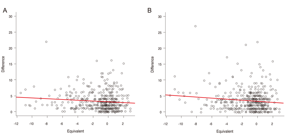
Fig. 1 Plot diagrams of temporal (A) and clock-hour 9 (B) with spherical equivalent. Positive correlations between the standard deviation in retinal nerve fiber layer thickness at clock-hour 9 and spherical equivalent.
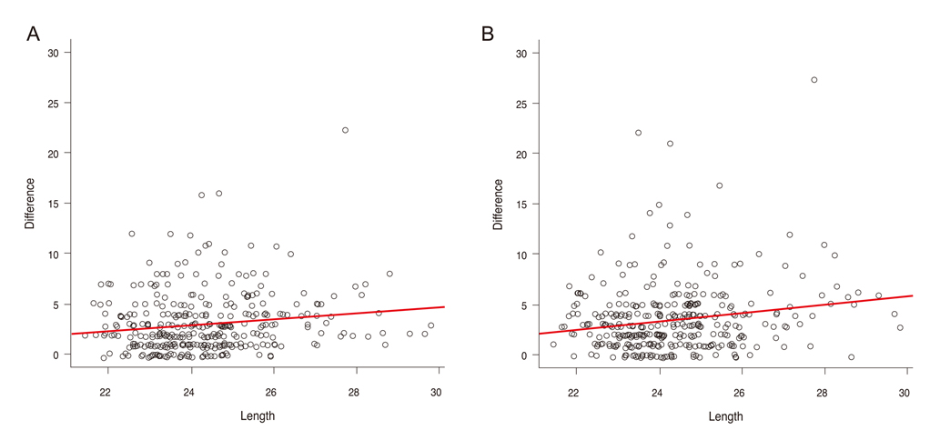
Fig. 2 Plot diagrams of temporal (A) and clock-hour 9 (B) with axial length. Positive correlations were observed between the standard deviation in retinal nerve fiber layer thickness at clock-hour 9 and axial length.
Reference
-
1. Vizzeri G, Weinreb RN, Gonzalez-Garcia AO, et al. Agreement between spectral-domain and time-domain OCT for measuring RNFL thickness. Br J Ophthalmol. 2009. 93:775–781.2. Seet B, Wong TY, Tan DT, et al. Myopia in Singapore: taking a public health approach. Br J Ophthalmol. 2001. 85:521–526.3. Lin LL, Shih YF, Hsiao CK, et al. Epidemiologic study of the prevalence and severity of myopia among schoolchildren in Taiwan in 2000. J Formos Med Assoc. 2001. 100:684–691.4. Suzuki Y, Iwase A, Araie M, et al. Risk factors for open-angle glaucoma in a Japanese population: the Tajimi Study. Ophthalmology. 2006. 113:1613–1617.5. Perkins ES, Phelps CD. Open angle glaucoma, ocular hypertension, low-tension glaucoma, and refraction. Arch Ophthalmol. 1982. 100:1464–1467.6. Grodum K, Heijl A, Bengtsson B. Refractive error and glaucoma. Acta Ophthalmol Scand. 2001. 79:560–566.7. Fong DS, Epstein DL, Allingham RR. Glaucoma and myopia: are they related? Int Ophthalmol Clin. 1990. 30:215–218.8. Seddon JM, Schwartz B, Flowerdew G. Case-control study of ocular hypertension. Arch Ophthalmol. 1983. 101:891–894.9. Leung CK, Chan WM, Yung WH, et al. Comparison of macular and peripapillary measurements for the detection of glaucoma: an optical coherence tomography study. Ophthalmology. 2005. 112:391–400.10. Hoffmann EM, Medeiros FA, Sample PA, et al. Relationship between patterns of visual field loss and retinal nerve fiber layer thickness measurements. Am J Ophthalmol. 2006. 141:463–471.11. Leung CK, Mohamed S, Leung KS, et al. Retinal nerve fiber layer measurements in myopia: an optical coherence tomography study. Invest Ophthalmol Vis Sci. 2006. 47:5171–5176.12. Choi SW, Lee SJ. Thickness changes in the fovea and peripapillary retinal nerve fiber layer depend on the degree of myopia. Korean J Ophthalmol. 2006. 20:215–219.13. Hoh ST, Lim MC, Seah SK, et al. Peripapillary retinal nerve fiber layer thickness variations with myopia. Ophthalmology. 2006. 113:773–777.14. Budenz DL, Chang RT, Huang X, et al. Reproducibility of retinal nerve fiber thickness measurements using the stratus OCT in normal and glaucomatous eyes. Invest Ophthalmol Vis Sci. 2005. 46:2440–2443.15. Budenz DL, Fredette MJ, Feuer WJ, Anderson DR. Reproducibility of peripapillary retinal nerve fiber thickness measurements with stratus OCT in glaucomatous eyes. Ophthalmology. 2008. 115:661–666.e4.16. Mayama C, Suzuki Y, Araie M, et al. Myopia and advanced-stage open-angle glaucoma. Ophthalmology. 2002. 109:2072–2077.17. Araie M, Arai M, Koseki N, Suzuki Y. Influence of myopic refraction on visual field defects in normal tension and primary open angle glaucoma. Jpn J Ophthalmol. 1995. 39:60–64.18. Greve EL, Furuno F. Myopia and glaucoma. Albrecht Von Graefes Arch Klin Exp Ophthalmol. 1980. 213:33–41.19. Park SH, Park KH, Kim JM, Choi CY. Relation between axial length and ocular parameters. Ophthalmologica. 2010. 224:188–193.20. Ray R, Stinnett SS, Jaffe GJ. Evaluation of image artifact produced by optical coherence tomography of retinal pathology. Am J Ophthalmol. 2005. 139:18–29.21. El-Ashry M, Appaswamy S, Deokule S, Pagliarini S. The effect of phacoemulsification cataract surgery on the measurement of retinal nerve fiber layer thickness using optical coherence tomography. Curr Eye Res. 2006. 31:409–413.22. Youm DJ, Kim JM, Park KH, Choi CY. The effect of soft contact lenses during the measurement of retinal nerve fiber layer thickness using optical coherence tomography. Curr Eye Res. 2009. 34:78–83.
- Full Text Links
-
- Actions
-
Cited
- CITED
-
- Close
- Share
-
- Similar articles
-
- Changes of Peripapillary Retinal Nerve Fiber Layer Thickness Profile According to Aging in Myopic Eyes
- Comparison of Time Domain OCT and Spectrum Domain OCT for Retinal Nerve Fiber Layer Assessment
- The Relationship among Refractive Power, Axial Length and Choroidal Thickness Measured by SD-OCT in Myopia
- Comparison of Retinal Nerve Fiber Layer Thickness Measured by Spectral-Domain and Time-Domain Optical Coherence Tomography
- Comparison of Diagnostic Ability of 3D and Stratus Optical Coherence Tomography in Early Glaucoma
