Measurement of Choroidal Thickness in Normal Eyes Using 3D OCT-1000 Spectral Domain Optical Coherence Tomography
- Affiliations
-
- 1Department of Ophthalmology, Hanyang University College of Medicine, Seoul, Korea. brlee@hanyang.ac.kr
- KMID: 1387391
- DOI: http://doi.org/10.3341/kjo.2012.26.4.255
Abstract
- PURPOSE
To study choroidal thickness and its topographic profile in normal eyes using 3D OCT-1000 spectral domain optical coherence tomography and the correlation with age and refractive error.
METHODS
Fifty-seven eyes (45 individuals) with no visual complaints or ocular disease underwent horizontal and vertical line scanning using 3D OCT-1000. The definition of choroidal thickness was the vertical distance between the posterior edge of the hyper-reflective retinal pigment epithelium and the choroid/sclera junction. Choroidal thickness was measured in the subfoveal area at 500 microm intervals from the fovea to 2,500 microm in the nasal, temporal, superior, and inferior regions. The spherical equivalent refractive error was measured by autorefractometry. Statistical analysis was used to confirm the correlations of choroidal thickness with age and refraction error.
RESULTS
The mean age of the 45 participants (57 eyes) was 45.28 years. Detailed visualization of the choroid for measuring its thickness was possible in 63.3% of eyes. The mean subfoveal choroidal thickness was found to be 270.8 microm (standard deviation [SD], +/-51 microm), in horizontal scanning and 275.0 microm (SD, +/-49 microm) in vertical scanning. The temporal choroidal thickness was greater than any 500 microm interval in corresponding locations, and there was no significant difference between the superior and inferior choroid as far as 2,000 microm from the fovea. Age and refractive error were associated with subfoveal choroidal thickness in terms of regression (p < 0.05).
CONCLUSIONS
Choroidal thickness in normal Korean eyes can be measured using 3D OCT-1000 with high resolution line scanning. The topographical profile of choroidal thickness varies depending on its location. Age and refractive error are essential factors for interpretation of choroidal thickness.
MeSH Terms
Figure
-
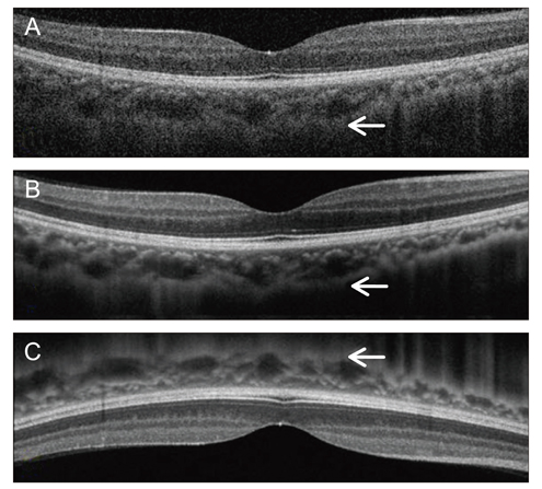
Fig. 1 Choroidal B-scan image without averaging in the conventional method shows an obscure choroid/sclera junction (A). Averaging of 50 B-scan images on choroidal mode improves choroidal visualization, but a partially unclear choroid/sclera junction image is produced (B). The device was pushed closely to the eye to obtain an inverted image. Using this technique, the zero-delay line that had highest sensitivity was closer to the choroid, and more accurate image acquisition was possible (compare arrows) (C).
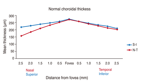
Fig. 2 Mean choroidal thickness at each of 22 locations in healthy Korean individuals. Subfoveal choroidal thickness is significantly greater than any other locations (paired t-test, p < 0.05, respectively). There is no statistically significant difference between horizontal and vertical subfoveal choroidal thickness (paired t-test, p = 0.131). S = superior; I = inferior; N = nasal; T = temporal.
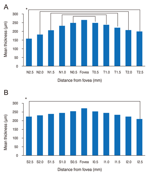
Fig. 3 (A) Temporal choroid is thicker at any corresponding location than the nasal choroid in horizontal scan (paired t-test, p < 0.05, respectively). (B) Superior choroid at 2,500 µm from the fovea is thicker than the inferior choroid (paired t-test, p < 0.05), and there is no difference in the vertical scan. *p < 0.05. S = superior; I = inferior; N = nasal; T = temporal.
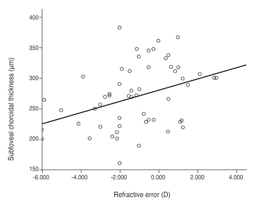
Fig. 4 Relationship between refractive error and subfoveal choroidal thickness. The plot shows a significant positive correlation in simple linear regression analysis (p = 0.046, y = 7.62x + 274.13, R2 = 0.101). D = diopters.
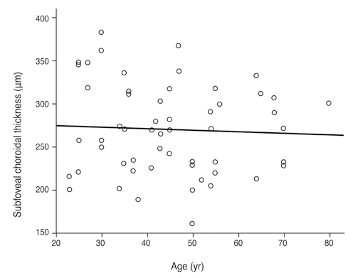
Fig. 5 Relationship between age and subfoveal choroidal thickness. Age is not related to choroidal thickness in simple linear regression (p = 0.688, y = -0.188x + 279.35, R2 = 0.003). Age is negatively correlated with choroidal thickness in multiple linear regression (p = 0.015, y = 341.584 - 1.306x1 + 13.621x2, R2 = 0.202).
Reference
-
1. Ikuno Y, Kawaguchi K, Nouchi T, Yasuno Y. Choroidal thickness in healthy Japanese subjects. Invest Ophthalmol Vis Sci. 2010. 51:2173–2176.2. Spaide RF, Koizumi H, Pozzoni MC. Enhanced depth imaging spectral-domain optical coherence tomography. Am J Ophthalmol. 2008. 146:496–500.3. Margolis R, Spaide RF. A pilot study of enhanced depth imaging optical coherence tomography of the choroid in normal eyes. Am J Ophthalmol. 2009. 147:811–815.4. Manjunath V, Taha M, Fujimoto JG, Duker JS. Choroidal thickness in normal eyes measured using Cirrus HD optical coherence tomography. Am J Ophthalmol. 2010. 150:325–329.e1.5. Unterhuber A, Povazay B, Hermann B, et al. In vivo retinal optical coherence tomography at 1040 nm - enhanced penetration into the choroid. Opt Express. 2005. 13:3252–3258.6. Ikuno Y, Tano Y. Retinal and choroidal biometry in highly myopic eyes with spectral-domain optical coherence tomography. Invest Ophthalmol Vis Sci. 2009. 50:3876–3880.7. Kiel JW, van Heuven WA. Ocular perfusion pressure and choroidal blood flow in the rabbit. Invest Ophthalmol Vis Sci. 1995. 36:579–585.8. Ramrattan RS, van der Schaft TL, Mooy CM, et al. Morphometric analysis of Bruch's membrane, the choriocapillaris, and the choroid in aging. Invest Ophthalmol Vis Sci. 1994. 35:2857–2864.9. Feeney-Burns L, Burns RP, Gao CL. Age-related macular changes in humans over 90 years old. Am J Ophthalmol. 1990. 109:265–278.10. Sarks SH. Ageing and degeneration in the macular region: a clinico-pathological study. Br J Ophthalmol. 1976. 60:324–341.11. Maruko I, Iida T, Sugano Y, et al. Subfoveal choroidal thickness after treatment of central serous chorioretinopathy. Ophthalmology. 2010. 117:1792–1799.12. Imamura Y, Fujiwara T, Margolis R, Spaide RF. Enhanced depth imaging optical coherence tomography of the choroid in central serous chorioretinopathy. Retina. 2009. 29:1469–1473.13. Spaide RF. Enhanced depth imaging optical coherence tomography of retinal pigment epithelial detachment in age-related macular degeneration. Am J Ophthalmol. 2009. 147:644–652.14. Spaide RF. Age-related choroidal atrophy. Am J Ophthalmol. 2009. 147:801–810.15. Fujiwara T, Imamura Y, Margolis R, et al. Enhanced depth imaging optical coherence tomography of the choroid in highly myopic eyes. Am J Ophthalmol. 2009. 148:445–450.16. Reibaldi M, Boscia F, Avitabile T, et al. Enhanced depth imaging optical coherence tomography of the choroid in idiopathic macular hole: a cross-sectional prospective study. Am J Ophthalmol. 2011. 151:112–117.e2.
- Full Text Links
-
- Actions
-
Cited
- CITED
-
- Close
- Share
-
- Similar articles
-
- The Relationship among Refractive Power, Axial Length and Choroidal Thickness Measured by SD-OCT in Myopia
- Choroidal Thickness in Primary Open-Angle Glaucoma Using Spectral-Domain Optical Coherence Tomography
- Choroidal Thickness at the Outside of Fovea in Diabetic Retinopathy Using Spectral-Domain Optical Coherence Tomography
- Comparison of Retinal Nerve Fiber Layer Thickness Measured by Spectral-Domain and Time-Domain Optical Coherence Tomography
- Measurement of Deep Optic Nerve Complex Structures with Two Spectral Domain Optical Coherence Tomography Instruments
