Ann Lab Med.
2012 May;32(3):177-183. 10.3343/alm.2012.32.3.177.
Changes in Urinary Stone Composition in the Tunisian Population: A Retrospective Study of 1,301 Cases
- Affiliations
-
- 1Department of Biochemistry and Toxicology, University Hospital, Monastir, Tunisia. akram_alaya@yahoo.co.uk
- 2Department of Pediatric Surgery, University Hospital, Monastir, Tunisia.
- 3Department of Urology, University Hospital, Monastir, Tunisia.
- KMID: 1381681
- DOI: http://doi.org/10.3343/alm.2012.32.3.177
Abstract
- BACKGROUND
Studies that evaluate the effect of age on stone composition are scarce. The aim of this study was to highlight the changes in epidemiological characteristics (stone composition and location) of urolithiasis according to patients' age.
METHODS
We studied 1,301 urolithiasis patients with age ranging from 6 months to 92 yr (781 males and 520 females). Stone analysis was performed using a stereomicroscope and infrared spectroscopy to determine the morphological type and molecular composition of each stone.
RESULTS
The annual average incidence of new stone formation was 31.7 per 100,000 persons. In 71.8% of cases, calculi were located in the upper urinary tract. Compared to other age groups, children and old men were more affected by bladder stones. Calcium oxalate monohydrate was the most frequent stone component, even though its frequency decreased with age (59.5% in young adults and 43.7% in the elderly, P<0.05) in favor of an increase in uric acid stones (11.5% in young adults and 36.4% in the elderly, P<0.05). Struvite stones were rare (3.8%) and more frequent in children than in adults.
CONCLUSIONS
The analysis of these data showed that urinary stones in Tunisian patients are tending to evolve in the same direction as the stones in patients from industrialized countries.
Keyword
MeSH Terms
-
Adolescent
Adult
Age Factors
Aged
Aged, 80 and over
Calcium Oxalate/chemistry
Child
Child, Preschool
Female
Humans
Infant
Kidney Calculi/chemistry/diagnosis/epidemiology
Magnesium Compounds/chemistry
Male
Middle Aged
Phosphates/chemistry
Retrospective Studies
Spectrophotometry, Infrared
Tunisia/epidemiology
Uric Acid/chemistry
Urinary Bladder Calculi/chemistry/diagnosis/epidemiology
Urinary Calculi/*chemistry/diagnosis/epidemiology
Young Adult
Figure
-
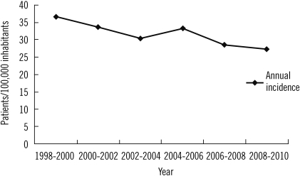
Fig. 1 Changes in the annual incidence of urolithiasis throughout the study period.
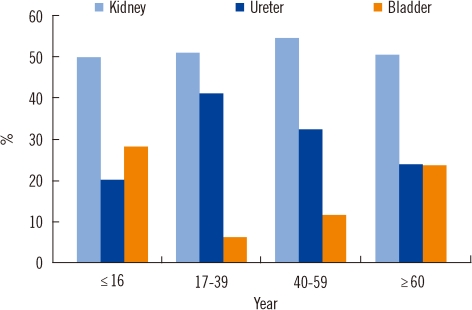
Fig. 2 Changes in stone location according to age (N=1,301).
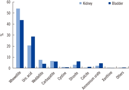
Fig. 3 Differences in kidney and bladder stone composition.
Cited by 1 articles
-
Analysis of Urinary Stone Composition: A Retrospective Single Center Study during the Last Five Years (2009-2013)
Pil Moon Kang, Won Ik Seo, Dong Il Kang
Korean J Urogenit Tract Infect Inflamm. 2014;9(1):44-49. doi: 10.14777/kjutii.2014.9.1.44.
Reference
-
1. Trinchieri A. Epidemiology of urolithiasis. Arch Ital Urol Androl. 1996; 68:203–249. PMID: 8936716.2. Ramello A, Vitale C, Marangella M. Epidemiology of nephrolithiasis. J Nephrol. 2000; 13(Suppl 3):S45–S50. PMID: 11132032.3. Robertson WG, Hughes H. Ryall R, Bais R, editors. Epidemiology of urinary stone disease in Saudi Arabia. Urolithiasis 2. 1994. New York London: Plenum Press;p. 453–455.
Article4. El-Reshaid K, Mughal H, Kapoor M. Epidemiological profile, mineral metabolic pattern and crystallographic analysis of urolithiasis in Kuwait. Eur J Epidemiol. 1997; 13:229–234. PMID: 9085010.5. Costa-Bauzá A, Ramis M, Montesinos V, Grases F, Conte A, Pizá P, et al. Type of renal calculi: variation with age and sex. World J Urol. 2007; 25:415–421. PMID: 17525848.
Article6. Alaya A, Najjar MF, Nouri A. Changes in stone composition according to age in Tunisian pediatric patients. Int Urol Nephrol. 2010; 42:621–628. PMID: 19937117.
Article7. Daudon M, Doré JC, Jungers P, Lacour B. Changes in stone composition according to age and gender of patients: a multivariate epidemiological approach. Urol Res. 2004; 32:241–247. PMID: 15127165.
Article8. Koide T, Itatani H, Yoshioka T, Ito H, Namiki M, Nakano E, et al. Clinical manifestations of calcium oxalate monohydrate and dihydrate urolithiasis. J Urol. 1982; 127:1067–1069. PMID: 7087010.
Article9. Daudon M, Donsimoni R, Hennequin C, Fellahi S, Le Moel G, Paris M, et al. Sex- and age-related composition of 10 617 calculi analyzed by infrared spectroscopy. Urol Res. 1995; 23:319–326. PMID: 8839389.
Article10. López M, Hoppe B. History, epidemiology and regional diversities of urolithiasis. Pediatr Nephrol. 2010; 25:49–59. PMID: 21476230.
Article11. Elder JS. Behrman RE, Kliegman RM, editors. Urinary lithiasis. Nelson textbook of pediatrics. 2004. 17th ed. Philadelphia: Saunders;p. 1822–1826.
Article12. Oner A, Demircin G, Ipekçioğlu H, Bülbül M, Ecin N. Etiological and clinical patterns of urolithiasis in Turkish children. Eur Urol. 1997; 31:453–458. PMID: 9187907.
Article13. Power C, Barker DJ, Blacklock NJ. Incidence of renal stones in 18 British towns. A collaborative study. Br J Urol. 1987; 59:105–110. PMID: 3828703.
Article14. Ahlstrand C, Tiselius HG. Renal stone disease in a Swedish district during one year. Scand J Urol Nephrol. 1981; 15:143–146. PMID: 7330608.
Article15. Serio A, Fraioli A. Epidemiology of nephrolithiasis. Nephron. 1999; 81(S1):S26–S30.
Article16. Curhan GC, Rimm EB, Willett WC, Stampfer MJ. Regional variation in nephrolithiasis incidence and prevelance among United States men. J Urol. 1994; 151:838–841. PMID: 8126805.17. Neuzillet Y, Lechevallier E, Ballanger P, Ferriere JM, Saussine C, Doré B, et al. Urinary stones in subjects over the age of sixty. Prog Urol. 2004; 14:479–484. PMID: 15776895.18. Daudon M. Évolution de la composition et de la localisation des calculs chez le sujet âgé. Feuillets de Biologie. 2003; 25:51–54.19. El-Reshaid K, Mughal H, Kapoor M. Epidemiological profile, mineral metabolic pattern and crystallographic analysis of urolithiasis in Kuwait. Eur J Epidemiol. 1997; 13:229–234. PMID: 9085010.20. Daudon M. Comment analyser un calcul et comment interpréter le résultat. L' Eurobiologiste. 1993; 203:35–46.21. Daudon M, Bounxouei B, Santa Cruz F, Leite da Silva S, Diouf B, Angwafoo FF 3rd, et al. Composition of renal stones currently observed in non-industrialized countries. Prog Urol. 2004; 14:1151–1161. PMID: 15751409.22. Silva SF, Matos DC, Silva SL, Daher Ede F, Campos Hde H, Silva CA. Chemical and morphological analysis of kidney stones: a double-blind comparative study. Acta Cir Bras. 2010; 25:444–448. PMID: 20877956.
Article23. Stitchantrakul W, Kochakarn W, Ruangraksa C, Domrongkitchaiporn S. Urinary risk factors for recurrent calcium stone formation in Thai stone formers. J Med Assoc Thai. 2007; 90:688–698. PMID: 17487123.24. Baker PW, Coyle P, Bais R, Rofe AM. Influence of season, age, and sex on renal stone formation in South Australia. Med J Aust. 1993; 159:390–392. PMID: 8377690.
Article25. Djelloul Z, Djelloul A, Bedjaoui A, Kaid-Omar Z, Attar A, Daudon M, et al. Urinary stones in Western Algeria: study of the composition of 1,354 urinary stones in relation to their anatomical site and the age and gender of the patients. Prog Urol. 2006; 16:328–335. PMID: 16821346.26. Abrams SA. Calcium turnover and nutrition through the life cycle. Proc Nutr Soc. 2001; 60:283–289. PMID: 11681644.27. Taylor EN, Stampfer MJ, Curhan GC. Dietary factors and the risk of incident kidney stones in men: new insights after 14 years of follow-up. J Am Soc Nephrol. 2004; 15:3225–3232. PMID: 15579526.
Article28. Leusmann DB, Blaschke R, Schmandt W. Results of 5,035 stone analyses: a contribution to epidemiology of urinary stone disease. Scand J Urol Nephrol. 1990; 24:205–210. PMID: 2237297.
Article29. Sun X, Shen L, Cong X, Zhu H, He L, Lu J. Infrared spectroscopic analysis of 5,248 urinary stones from Chinese patients presenting with the first stone episode. Urol Res. 2011; 39:339–343. PMID: 21249491.
Article30. Najjar MF, Najjar F, Boukef K, Oueslati A, Memmi J, Bechraoui T. La lithiase infantile dans la région de Monastir étude clinique et biologique. Le Biologiste. 1986; 165:31–39.31. Strohmaier WL, Weigl A. Jungers P, Daudon M, editors. Stone composition in upper Franconia-unusually high percentage of uric acid lithiasis. Renal stone disease. 1997. Amsterdam: Elsevier Science;p. 10–11.32. Curhan GC, Willett WC, Speizer FE, Spiegelman D, Stampfer MJ. Comparison of dietary calcium with supplemental calcium and other nutrients as factors affecting the risk for kidney stones in women. Ann Intern Med. 1997; 126:497–504. PMID: 9092314.
Article33. Grases F, Costa-Bauza A, Prieto RM. Renal lithiasis and nutrition. Nutr J. 2006; 5:23. PMID: 16956397.
Article34. Alaya A, Nouri A, Najjar MF. Urinary stone composition in pediatric patients: a retrospective study of 205 cases. Clin Chem Lab Med. 2011; 49:243–248. PMID: 21143010.
Article35. Agarwal BN, Cabebe FG. Renal acidification in elderly subjects. Nephron. 1980; 26:291–295. PMID: 7453916.
Article36. Daudon M, Jungers P. Diabetes and calculations. Feuillets de Biologie. 2001; 239:37–39.37. Siener R, Glatz S, Nicolay C, Hesse A. The role of overweight and obesity in calcium oxalate stone formation. Obes Res. 2004; 12:106–113. PMID: 14742848.
Article38. Trinchieri A, Ostini F, Nespoli R, Rovera F, Montanari E, Zanetti G. A prospective study of recurrence rate and risk factors for recurrence after a first renal stone. J Urol. 1999; 162:27–30. PMID: 10379732.
Article39. Siener R, Glatz S, Nicolay C, Hesse A. Prospective study on the efficacy of a selective treatment and risk factors for relapse in recurrent calcium oxalate stone patients. Eur Urol. 2003; 44:467–474. PMID: 14499683.
Article40. Koşar A, Sarica K, Aydos K, Küpeli S, Türkölmez K, Göğüs O. Comparative study of long-term stone recurrence after extracorporeal shock wave lithotripsy and open stone surgery for kidney stones. Int J Urol. 1999; 6:125–129. PMID: 10226822.
- Full Text Links
-
- Actions
-
Cited
- CITED
-
- Close
- Share
-
- Similar articles
-
- The Predictability of Stone Composition According to the Radiodensity of Urinary Stone on KUB Film
- The study on Non-successful Cases of ESWL in the Upper Urinary Tract Stone using Wolf Piezolith 2300 Lithotriptor(R)
- Spectroscopic Studies of Urinary Stone
- Biochemical Characteristics of Serum and Urine in the Patients with Uric Acid Stone
- The Values of Calcium, Uric Acid, Magnesium and Magnesium/Calcium Ratio in Urine with the Urinary Calculi
