Sertoli cell tumors associated with feminizing syndrome and spermatic cord torsion in two cryptorchid dogs
- Affiliations
-
- 1Department of Veterinary Public Health, University of Messina, 98168 Messina, Italy. marinog@unime.it
- KMID: 1376197
- DOI: http://doi.org/10.4142/jvs.2012.13.2.207
Abstract
- The association of cryptorchidism, functional Sertoli cell tumors, and spermatic cord torsion has been rarely reported in the literature. Two dogs were admitted for bilateral skin alopecia and weight loss. Both animals were cryptorchid and displayed a pendulous preputial sheath, prostate hypertrophy, and increased levels of circulating oestrogen. Transabdominal palpation and ultrasonography revealed the presence of neoplastic retained gonads. During surgery, spermatic cord torsion was also detected in the enlarged neoplastic testes of both dogs. Histologic examination confirmed the presence of Sertoli cell tumors that were primarily responsible for the feminizing syndrome. Complete remission of all symptoms occurred within 3 months after orchiectomy.
MeSH Terms
Figure
-
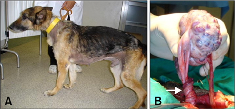
Fig. 1 Case 1. (A) A dog presenting characteristic bilateral symmetrical alopecia with hyperkeratosis involving the ventral abdomen, flanks, thorax, and neck. (B) The left testis at surgery was greatly increased in size and had multiple spermatic cord torsions (arrow).
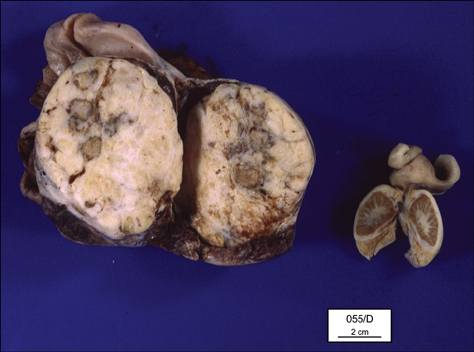
Fig. 2 Case 2. Gross appearance of a totally neoplastic testis compared to the contralateral atrophic one.
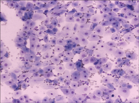
Fig. 3 Case 2. Epithelial cells with signs of squamous metaplasia collected from a prostatic cyst. May-Grünwald-Giemsa stain, ×250.
Cited by 1 articles
-
Unilateral cryptorchidism induces morphological changes of testes and hyperplasia of Sertoli cells in a dog
Joon Ho Moon, Dae Young Yoo, Young Kwang Jo, Geon A Kim, Hyo Young Jung, Jung Hoon Choi, In Koo Hwang, Goo Jang
Lab Anim Res. 2014;30(4):185-189. doi: 10.5625/lar.2014.30.4.185.
Reference
-
1. Feldman EC, Nelson RW, editors. Canine and Feline Endocrinology and Reproduction. 1987. 2nd ed. Philadelphia: Saunders;481–523.2. Gill CW. Prostatic adenocarcinoma with concurrent Sertoli cell tumor in a dog. Can Vet J. 1981. 22:230–233.3. Hayes HM Jr, Wilson GP, Pendergrass TW, Cox VS. Canine cryptorchism and subsequent testicular neoplasia: case-control study with epidemiologic update. Teratology. 1985. 32:51–56.
Article4. Heidbrink U, Kaup FJ. Case report: Sertoli cell tumor with feminisation syndrome in a male dog (Deutch Langhaar). Kleintierpraxis. 1990. 35:661–665.5. Laing EJ, Harari J, Smith CW. Spermatic cord torsion and Sertoli cell tumor in a dog. J Am Vet Med Assoc. 1983. 183:879–881.6. Metzger FL, Hattel AL, White DG. Hematuria, hyperestrogenemia, and hyperprogesteronemia due to a Sertoli-cell tumor in a bilaterally cryptorchid dog. Canine Pract. 1993. 18:32–35.7. Miyabayashi T, Biller DS, Cooley AJ. Ultrasonographic appearance of torsion of a testicular seminoma in a cryptorchid dog. J Small Anim Pract. 1990. 31:401–403.
Article8. Pearson H, Kelly DF. Testicular torsion in the dog: a review of 13 cases. Vet Rec. 1975. 97:200–204.
Article9. Reif JS, Brodey RS. The relationship between cryptorchidism and canine testicular neoplasia. J Am Vet Med Assoc. 1969. 155:2005–2010.10. Sanpera N, Masot N, Janer M, Romeo C, de Pedro R. Oestrogen-induced bone marrow aplasia in a dog with a Sertoli cell tumour. J Small Anim Pract. 2002. 43:365–369.
Article11. Spackman CJ, Roth L. Prostatic cyst and concurrent Sertoli cell tumor in a dog. J Am Vet Med Assoc. 1988. 192:1096–1098.
