Sequential Intrastromal Corneal Ring Implantation and Cataract Surgery in a Severe Keratoconus Patient with Cataract
- Affiliations
-
- 1SU Yonsei Eye Clinic, Seoul, Korea. grace04@freechal.com
- KMID: 1376131
- DOI: http://doi.org/10.3341/kjo.2012.26.3.226
Abstract
- A 49-year-old man with an uncorrected visual acuity (UCVA) of 20 / 1000, a best spectacle-corrected visual acuity (BSCVA) of 20 / 400, keratometric readings of K1 = 59.88 x 82degrees / K2 = 45.88 x 172degrees, and an inferior steepening that was consistent with keratoconus in his left eye was treated with clear-cornea phacoemulsification and an intraocular lens (IOL) implantation after insertion of keraring intrastromal corneal ring segments for severe keratoconus and cataract. An asymmetrical pair of kerarings was implanted with the assistance of a femtosecond laser in September 2008; the one segment was 250 microm and the other was 150 microm and both were placed at 70degrees. Three months after the kerarings were implanted, clear-cornea phacoemulsification and IOL implantation were performed on the left eye. After surgery, both the UCVA and the BSCVA of the left eye improved by eight lines. Postoperative central keratometry showed a decrease of 7.35 diopters in the left eye. Both the postoperative refraction (-0.75 -0.75 x 60degrees) and the keratometric reading (K1 = 50.05 x 93degrees / K2 = 48.83 x 3degrees) remained stable one month following the procedures. Thus, the sequential order of intrastromal corneal rings implantation and cataract surgery can be considered as a treatment option in patients with severe keratoconus and cataract.
Keyword
MeSH Terms
Figure
-
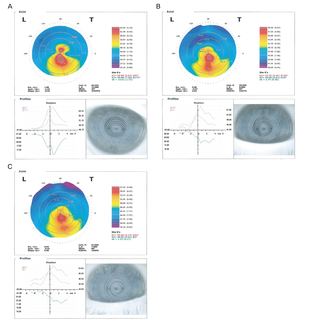
Fig. 1 Preoperative and postoperative topographies. (A) Preoperative topography shows a characteristic keratoconus with 60.04 diopter (D, central K) and 32,629 (KISA%). (B) Postoperative topography three months after implantation of the kerarings shows an improved corneal steepening with 55.04 D (central K). (C) Postoperative topography one month after the cataract surgery shows a 52.69 D in the central K. L = left eye; T = temporal; Ecc = eccentricity; Asph = asphericity; I_S = inferior superior dioptric asymmetricitiy; K = keratometric value; SRAX = skewed radial axis; dK = difference K (K2-K1).

Fig. 2 Postoperative photographs. (A) Postoperative photograph after implantation of the kerarings, which shows that the asymmetric keraring segments are well implanted at 70°. (B) Postoperative photograph after the cataract surgery shows a well implanted intraocular lens.
Reference
-
1. Rabinowitz YS. Keratoconus. Surv Ophthalmol. 1998. 42:297–319.2. Krachmer JH, Feder RS, Belin MW. Keratoconus and related noninflammatory corneal thinning disorders. Surv Ophthalmol. 1984. 28:293–322.3. Tomidokoro A, Oshika T, Amano S, et al. Changes in anterior and posterior corneal curvatures in keratoconus. Ophthalmology. 2000. 107:1328–1332.4. Antalis JJ, Lembach RG, Carney LG. A comparison of the TMS-1 and the corneal analysis system for the evaluation of abnormal corneas. CLAO J. 1993. 19:58–63.5. Thebpatiphat N, Hammersmith KM, Rapuano CJ, et al. Cataract surgery in keratoconus. Eye Contact Lens. 2007. 33:244–246.6. Colin J, Cochener B, Savary G, Malet F. Correcting keratoconus with intracorneal rings. J Cataract Refract Surg. 2000. 26:1117–1122.7. Alio JL, Shabayek MH. Intracorneal asymmetrical rings for keratoconus: where should the thicker segment be implanted? J Refract Surg. 2006. 22:307–309.8. Alio JL, Shabayek MH, Artola A. Intracorneal ring segments for keratoconus correction: long-term follow-up. J Cataract Refract Surg. 2006. 32:978–985.9. Burris TE, Ayer CT, Evensen DA, Davenport JM. Effects of intrastromal corneal ring size and thickness on corneal flattening in human eyes. Refract Corneal Surg. 1991. 7:46–50.10. Shabayek MH, Alio JL. Intrastromal corneal ring segment implantation by femtosecond laser for keratoconus correction. Ophthalmology. 2007. 114:1643–1652.11. Coskunseven E, Kymionis GD, Tsiklis NS, et al. One-year results of intrastromal corneal ring segment implantation (KeraRing) using femtosecond laser in patients with keratoconus. Am J Ophthalmol. 2008. 145:775–779.12. Coskunseven E, Kymionis GD, Talu H, et al. Intrastromal corneal ring segment implantation with the femtosecond laser in a post-keratoplasty patient with recurrent keratoconus. J Cataract Refract Surg. 2007. 33:1808–1810.13. Kwitko S, Severo NS. Ferrara intracorneal ring segments for keratoconus. J Cataract Refract Surg. 2004. 30:812–820.14. Alio JL, Artola A, Ruiz-Moreno JM, et al. Changes in keratoconic corneas after intracorneal ring segment explantation and reimplantation. Ophthalmology. 2004. 111:747–751.15. Rahman I, Carley F, Hillarby C, et al. Penetrating keratoplasty: indications, outcomes, and complications. Eye (Lond). 2009. 23:1288–1294.16. Patel SV, Hodge DO, Bourne WM. Corneal endothelium and postoperative outcomes 15 years after penetrating keratoplasty. Am J Ophthalmol. 2005. 139:311–319.
- Full Text Links
-
- Actions
-
Cited
- CITED
-
- Close
- Share
-
- Similar articles
-
- Effect of Sequential Intrastromal Corneal Ring Segment Implantation and Corneal Collagen Crosslinking in Corneal Ectasia
- Intrastromal Corneal Ring Segments (KeraRing(R)) Implantation for the Correction of Keratoconus
- Clinical Results of Intacs(R) Ring Implantation in Keratoconus or Keratectasia
- The Clinical Results of Intacs(R) Ring Implantation by Manual Tunnel Creation in Patients with Keratoconus
- Cataract Extraction after Penetrating Keratoplasty
