Unusual Involvement of IgG4-Related Sclerosing Disease in Lacrimal and Submandibular Glands and Extraocular Muscles
- Affiliations
-
- 1Department of Ophthalmology, Hanyang University College of Medicine, Seoul, Korea.
- 2Department of Pathology, Hanyang University College of Medicine, Seoul, Korea.
- 3Department of Ophthalmology, Gavin Herbert Eye Institute, University of California Irvine, Irvine, CA, USA. yoonjl2@uci.edu
- KMID: 1376129
- DOI: http://doi.org/10.3341/kjo.2012.26.3.216
Abstract
- Chronic sclerosing sialadenitis, also known as Kuttner tumor, is a chronic inflammatory disease of the salivary glands that is reported in a few cases in medical literature. Recent reports suggest that certain aspects of sclerosing diseases, including chronic sclerosing sialadenitis or dacryoadenitis, should be classified under immunoglobulin G4 (IgG4)-related sclerosing disease based on immunohistochemical studies. This study reports an unusual case of IgG4-related sclerosing disease appearing simultaneously in the lacrimal glands, submandibular glands, and extraocular muscles. A 56-year-old male presented with complaints of bilateral eyelid swelling and proptosis that began two years ago. Computed tomography confirmed that bilateral submandibular enlargements also existed five years ago in the subject. Orbital computed tomography and magnetic resonance imaging revealed bilateral lacrimal gland enlargement and thickening of extraocular muscles. Typical findings of chronic sclerosing dacryoadenitis were revealed upon pathologic exam of the right lacrimal gland. Immunostaining revealed numerous IgG4-positive plasma cells. Through these clinical features, we make a diagnosis of IgG4-relataed sclerosing disease in the subject.
MeSH Terms
-
Biopsy, Fine-Needle
Diagnosis, Differential
Facial Muscles/*immunology/pathology/radiography
Follow-Up Studies
Humans
Immunoglobulin G/*immunology/metabolism
Immunohistochemistry
Lacrimal Apparatus/*immunology/metabolism/pathology
Lacrimal Duct Obstruction/complications/diagnosis/*immunology
Magnetic Resonance Imaging
Male
Middle Aged
Sclerosis
Submandibular Gland/*immunology/pathology/radiography
Submandibular Gland Diseases/complications/diagnosis/*immunology
Tomography, X-Ray Computed
Figure
-
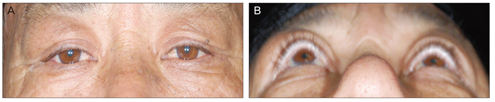
Fig. 1 Complaint from a 56-year-old male of bilateral eyelid swelling and proptosis that started 2 years ago. Symptoms were more promi nent in the right eye. (A) Frontal view and (B) inferior view.
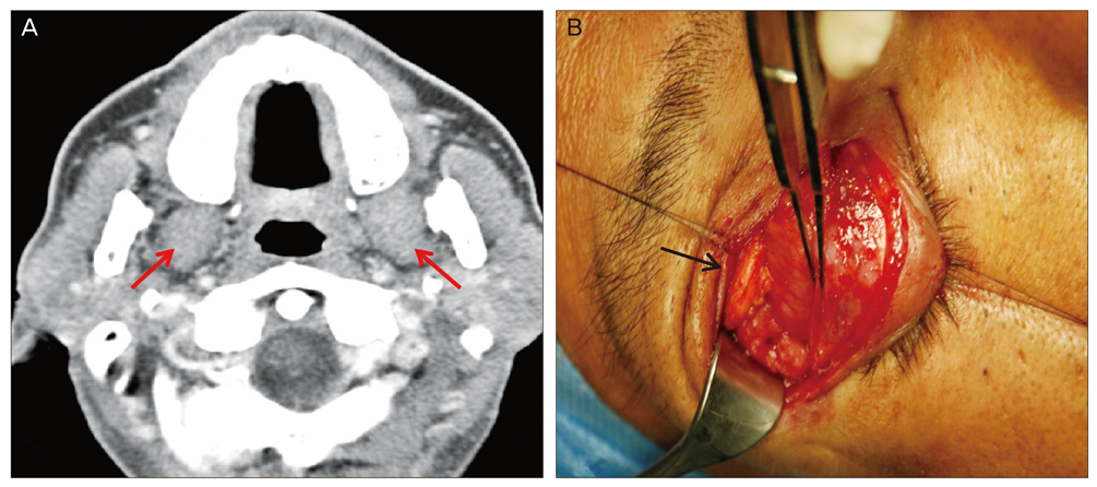
Fig. 2 (A) Both submandibular enlargements (arrows) existed 5 years ago. Currently there have been no definite changes of size. (B) Incisional biopsy of right lacrimal mass was performed. Enlargement of lacrimal gland was found (arrow).

Fig. 3 (A) Axial view of orbital computed tomography (CT). Arrows indicate bilateral lacrimal gland enlargement. (B) Axial view of orbital magnetic resonance imaging (MRI) also shows bilateral lacrimal gland enlargement (arrows). (C) Coronal view of orbital CT demonstrates the enlargement of bilateral lateral recti (arrows) and right inferior rectus. (D) Coronal view of orbital MRI. Dashed arrow indicates the hypertrophy of right inferior rectus.
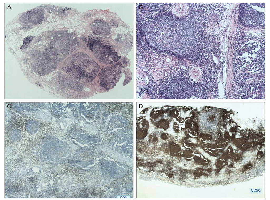
Fig. 4 (A,B) Light microscopic examination of tissue stained with H&E. (A) Lacrimal gland reveals dense lymphocyte hyperplasia and lymphoid follicles (×2). (B) Atrophic lacrimal ducts and periductal sclerosis are shown (×40). (C,D) Tissue with immunohistochemical stains. The hyperplastic lymphoid tissues are composed of polyclonal lymphoid cells, positive for CD 3 (C, ×40) and positive for CD 20 (D, ×2).
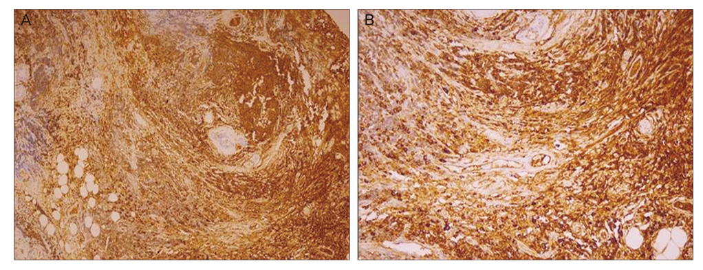
Fig. 5 (A,B) Immunostaining for immunoglobulin G4 (IgG4). Abundant IgG4-positive plasma cells (brown color) are demonstrated. Prevalence shows that Kuttner tumor is a kind of IgG4-related sclerosing disease (A, ×100; B, ×200).
Reference
-
1. Kuttner H. Uber entzundliche tumoren der submaxillar-speicheldruse. Bruns Beitr Klin Chir. 1896. 15:815–834.2. Williams HK, Connor R, Edmondson H. Chronic sclerosing sialadenitis of the submandibular and parotid glands: a report of a case and review of the literature. Oral Surg Oral Med Oral Pathol Oral Radiol Endod. 2000. 89:720–723.3. Blanco M, Mesko T, Cura M, Cabello-Inchausti B. Chronic sclerosing sialadenitis (Kuttner's tumor): unusual presentation with bilateral involvement of major and minor salivary glands. Ann Diagn Pathol. 2003. 7:25–30.4. Tiemann M, Teymoortash A, Schrader C, et al. Chronic sclerosing sialadenitis of the submandibular gland is mainly due to a T lymphocyte immune reaction. Mod Pathol. 2002. 15:845–852.5. Kitagawa S, Zen Y, Harada K, et al. Abundant IgG4-positive plasma cell infiltration characterizes chronic sclerosing sialadenitis (Kuttner's tumor). Am J Surg Pathol. 2005. 29:783–791.6. Cheuk W, Yuen HK, Chan JK. Chronic sclerosing dacryoadenitis: part of the spectrum of IgG4-related Sclerosing disease? Am J Surg Pathol. 2007. 31:643–645.7. Roh JL, Kim JM. Kuttner's tumor: unusual presentation with bilateral involvement of the lacrimal and submandibular glands. Acta Otolaryngol. 2005. 125:792–796.8. Jakobiec FA, Stacy RC, Mehta M, Fay A. IgG4-positive dacryoadenitis and Kuttner submandibular sclerosing inflammatory tumor. Arch Ophthalmol. 2010. 128:942–944.9. Lee LY, Chen TC, Kuo TT. Simultaneous occurrence of IgG4-related chronic sclerosing dacryoadenitis and chronic sclerosing sialadenitis associated with lymph node involvement and Warthin's tumor. Int J Surg Pathol. 2011. 19:369–372.10. Harrison JD, Epivatianos A, Bhatia SN. Role of microliths in the aetiology of chronic submandibular sialadenitis: a clinicopathological investigation of 154 cases. Histopathology. 1997. 31:237–251.11. Van der Zee JS, Aalberse RC. Immunochemical characteristics of IgG4 antibodies. N Engl Reg Allergy Proc. 1988. 9:31–33.12. Kamisawa T, Funata N, Hayashi Y, et al. A new clinicopathological entity of IgG4-related autoimmune disease. J Gastroenterol. 2003. 38:982–984.13. Kwon JE, Kim SK, Lee SR, et al. Chronic sclerosing dacryoadenitis: report of 2 cases. Korean J Pathol. 2008. 42:118–122.14. Yamamoto M, Takahashi H, Ohara M, et al. A new conceptualization for Mikulicz's disease as an IgG4-related plasmacytic disease. Mod Rheumatol. 2006. 16:335–340.15. Cheuk W, Yuen HK, Chan AC, et al. Ocular adnexal lymphoma associated with IgG4+ chronic sclerosing dacryoadenitis: a previously undescribed complication of IgG4-related sclerosing disease. Am J Surg Pathol. 2008. 32:1159–1167.16. Ochoa ER, Harris NL, Pilch BZ. Marginal zone B-cell lymphoma of the salivary gland arising in chronic sclerosing sialadenitis (Kuttner tumor). Am J Surg Pathol. 2001. 25:1546–1550.17. Sekine S, Nagata M, Watanabe T. Chronic sclerosing sialadenitis of the submandibular gland associated with idiopathic retroperitoneal fibrosis. Pathol Int. 1999. 49:663–667.18. Tsuneyama K, Saito K, Ruebner BH, et al. Immunological similarities between primary sclerosing cholangitis and chronic sclerosing sialadenitis: report of the overlapping of these two autoimmune diseases. Dig Dis Sci. 2000. 45:366–372.
- Full Text Links
-
- Actions
-
Cited
- CITED
-
- Close
- Share
-
- Similar articles
-
- A Case of IgG4-Related Sclerosing Disease Involving the Eyelid in an Idiopathic Sclerosing Myositis Patient
- Two Cases of Immunoglobulin G4-Related Sclerosing Disease in Submandibular Triangle
- IgG4-Related Sclerosing Sialadenitis: Report of Three Cases
- A Case of IgG4-Related Sclerosing Disease Involving the Optic Nerve
- Two Cases of Immunoglobulin G4-Related Sclerosing Disease Mimicking Nasopharyngeal Carcinoma
