Bell's Phenomenon during Screening Examination for Retinopathy of Prematurity
- Affiliations
-
- 1Department of Ophthalmology, Medical Research Institute, Pusan National University School of Medicine, Busan, Korea. hychoi@pusan.ac.kr
- 2Busan St. Mary's Medical Center, Busan, Korea.
- KMID: 1376124
- DOI: http://doi.org/10.3341/kjo.2012.26.3.189
Abstract
- PURPOSE
Bell's phenomenon (BP), which may disturb screening examinations for retinopathy of prematurity (ROP), is known to present infrequently in premature babies. Stress associated with the examinations can influence expression of BP. The authors of the present study evaluated BP during examinations for ROP.
METHODS
The present study included 102 eyes of 51 premature babies. Expression of BP was assessed at 3 steps of the examination in the following order: after insertion of a speculum, after illumination of an indirect ophthalmoscope and after scleral depression. The relationship between the expression of BP and the gestational age at the examination was analyzed in each step of the examination.
RESULTS
The frequency of BP after the speculum insertion and the illumination was 77% to 92% in infants 32 weeks of age or younger, and decreased significantly to 16% to 57% in infants 42 weeks of age or older (p < 0.005). BP after the scleral depression had no significant association with the gestational age. Frequency of BP increased significantly as the steps of the examination proceeded (p < 0.01).
CONCLUSIONS
BP was frequent in premature infants during ROP examination in spite of neurological immaturity. The examiner should take BP into consideration, which frequently occurs in younger infants.
MeSH Terms
-
Gestational Age
Humans
Incidence
Infant, Newborn
*Infant, Premature
Korea/epidemiology
Mass Screening/adverse effects/*methods/psychology
Ophthalmoscopy/*psychology
Retinopathy of Prematurity/*diagnosis/epidemiology
Retrospective Studies
Risk Factors
Stress, Psychological/*epidemiology/etiology
Vision Screening/adverse effects/*psychology
Figure
-
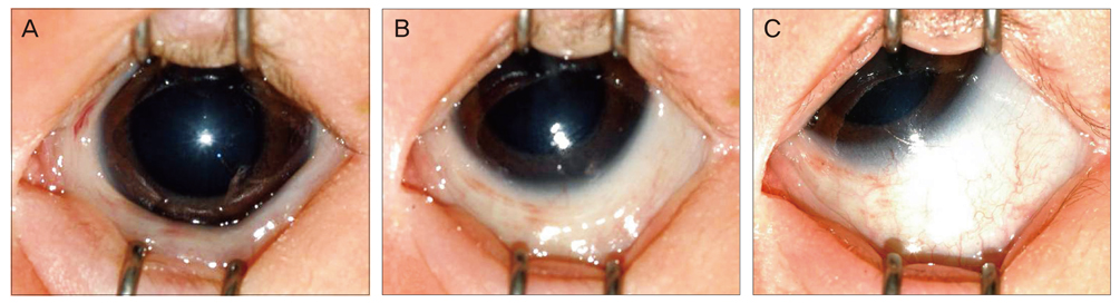
Fig. 1 Grading of Bell's phenomenon: (A) grade 0, no response; (B) grade 1, minimal response, or the center of cornea is seen; (C) grade 2, full response or the center of the cornea is not seen.
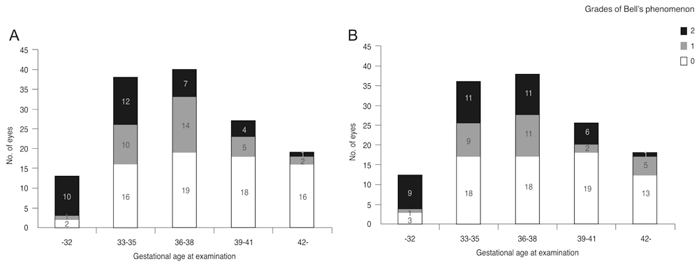
Fig. 2 Frequency and grade of Bell's phenomenon after insertion of speculum decrease significantly according to gestational age at examination for retinopathy of prematurity of (A) right eyes (p < 0.001) and (B) left eyes (p = 0.005).

Fig. 3 Frequency and grade of Bell's phenomenon after illumination of indirect ophthalmoscope decrease significantly according to gestational age at examination for retinopathy of prematurity of (A) right eyes (p < 0.001) and (B) left eyes (p = 0.001).

Fig. 4 Frequency and grade of Bell's phenomenon after scleral depression do not show a difference according to gestational age at examination for retinopathy of prematurity of (A) right eyes (p = 0.080) and (B) left eyes (p = 0.075).
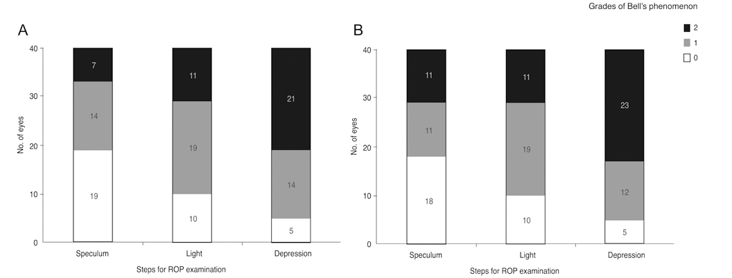
Fig. 5 Frequency and grade of Bell's phenomenon increase significantly according to progression of the examination in premature infants with a gestational age of 36 to 38 weeks at examination for retinopathy of prematurity (ROP) of (A) right eyes (p < 0.001) and (B) left eyes (p < 0.001). No difference was found between the right and the left eyes.
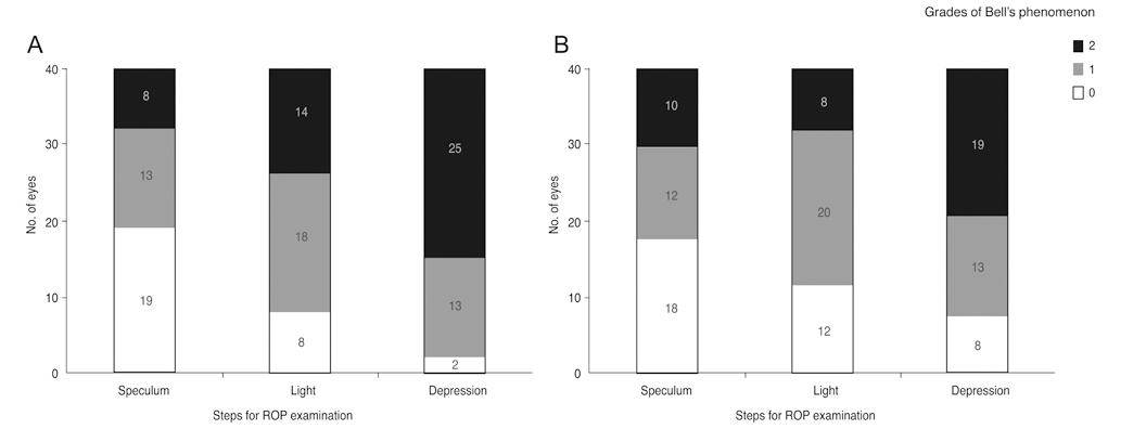
Fig. 6 Frequency and grade of Bell's phenomenon increase significantly according to progression of the examination in premature infants with a gestational age of 36 to 38 weeks at examination for retinopathy of prematurity (ROP) of (A) preceding eyes (p < 0.001) and (B) following eyes (p < 0.001). No difference was found between the preceding and the following eyes.
Reference
-
1. Bell C. On the motions of the eye, in illustration of the uses of the muscles and nerves of the orbit. Philos Trans R Soc Lond. 1823. 113:166–186.2. Hiraoka M. Physiological study of the Bell's phenomenon in human (author's transl). Nihon Ganka Gakkai Zasshi. 1979. 83:2184–2190.3. Ferrer JA. Conclusions from Bell's phenomenon variants. Trans Am Acad Ophthalmol Otolaryngol. 1973. 77:OP714–OP720.4. Francis IC, Loughhead JA. Bell's phenomenon. A study of 508 patients. Aust J Ophthalmol. 1984. 12:15–21.5. Snir M, Kremer I, Kuperman A, et al. Bell's phenomenon in newborns and premature babies. Br J Ophthalmol. 1996. 80:553–555.6. Park SW, Lee JE, Choi HY, Oum BS. Bell's phenomenon and conjunctival injury in screening examination for retinopathy of prematurity. J Korean Ophthalmol Soc. 2007. 48:1694–1698.7. Thorn F, Gwiazda J, Cruz AA, et al. The development of eye alignment, convergence, and sensory binocularity in young infants. Invest Ophthalmol Vis Sci. 1994. 35:544–553.8. Archer SM, Sondhi N, Helveston EM. Strabismus in infancy. Ophthalmology. 1989. 96:133–137.9. Paez JH, Isenberg S, Apt L. Torsion and elevation under general anesthesia and during voluntary eyelid closure (Bell phenomenon). J Pediatr Ophthalmol Strabismus. 1984. 21:22–24.
- Full Text Links
-
- Actions
-
Cited
- CITED
-
- Close
- Share
-
- Similar articles
-
- Bell's Phenomenon and Conjunctival Injury in Screening Examination for Retinopathy of Prematurity
- A Clinical Study of Retinopathy of Prematurity
- Screening Examination for Retinopathy of Prematurity with Dual Parameter Protocol
- The Effect of Cryotherapy and Laser Photocoagulation for the Retinopathy of Prematurity
- Effect of Cryotherapy for Retinopathy of Prematurity
