Morphological and Functional Correlates in Goldmann-Favre Syndrome: A Case Series
- Affiliations
-
- 1Elite School of Optometry, Sankara Nethralaya, Chennai, Tamil Nadu, India. madhavendra_opto@yahoo.co.in
- 2Shri Bhagwan Mahavir Vitreoretinal Services, Sankara Nethralaya, Chennai, Tamil Nadu, India.
- KMID: 1364870
- DOI: http://doi.org/10.3341/kjo.2012.26.2.143
Abstract
- The purpose of this study is to describe the correlation of findings between results from spectral domain optical coherence tomography (SD-OCT) and microperimetry in a case series regarding patients with Goldmann-Favre syndrome. Goldmann-Favre syndrome is a rare autosomal recessive hereditary vitreo-retinal degeneration that impacts the functionality of vision in subjects. Three men with this condition were assessed and subjected to microperimetry and SD-OCT. Two of the men were brothers. This study finds that the retinoschisis and macular cystoid changes noted in the SD-OCT matched the scotomas revealed by the microperimetry. The findings of each of the individual cases are reported herein.
Keyword
MeSH Terms
Figure
-
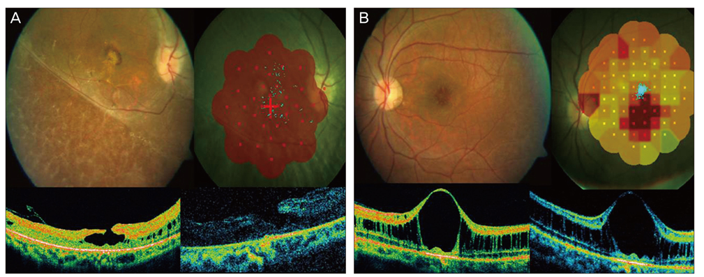
Fig. 1 (A) Fundus of the right eye shows lamellar macular holes with microcystic spaces and clumping of retinal pigment epithelium. Microperimetry shows grossly reduced retinal sensitivity. At the time of examination, time domain (Stratus) optical coherence tomography (OCT) showed confluent macular cystoid changes and foveal retinoschisis. Spectral domain OCT (SD-OCT) on the patient's follow-up visit revealed lamellar macular holes with macular schisis, microcystic spaces, and vitreomacular traction. (B) The left eye fundus shows a bicycle wheel pattern of foveal schisis. Microperimetry shows dense central scotomas. Images from time domain OCT and SD-OCT show cystic maculopathy with foveal schisis. SD-OCT images were taken at the time of the follow-up visit revealed cystoid macular oedema with inner layer schisis.
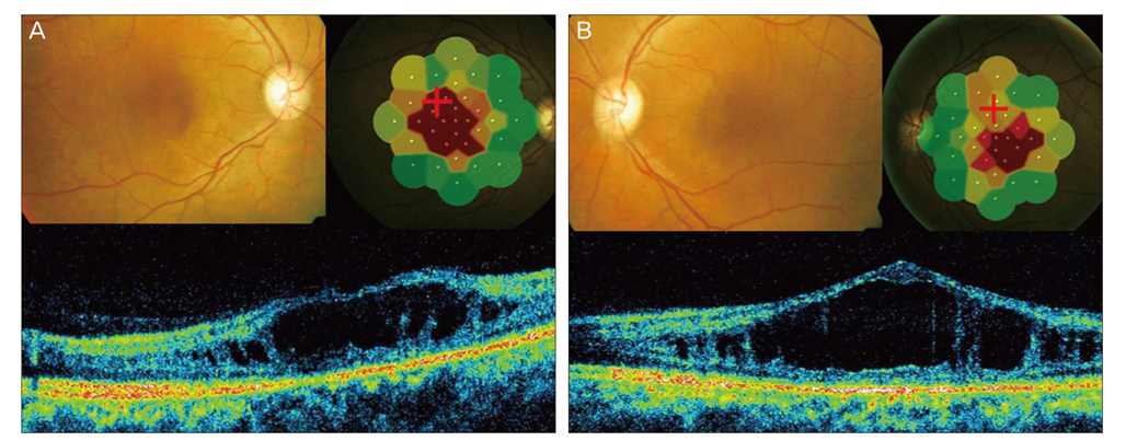
Fig. 2 (A) The right eye color fundus shows a bicycle wheel pattern in the fovea that is suggestive of foveal schisis with diffuse retinal pigment epithelium alterations. Microperimetry shows dense central scotoma with reduced sensitivity. Spectral domain optical coherence tomography (SD-OCT) shows foveal schisis with elevated foveal contour. (B) The left eye fundus shows a bicycle wheel pattern in the fovea that is suggestive of foveal schisis. Microperimetry shows reduced foveal sensitivity. SD-OCT imaging illustrates foveal schisis with elevated foveal contour.
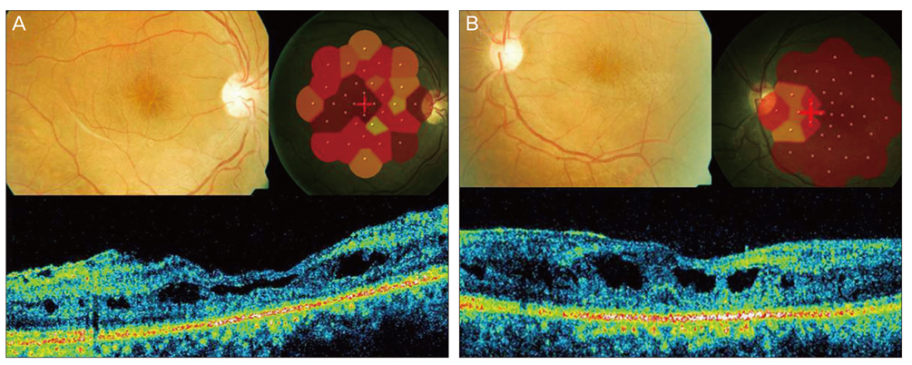
Fig. 3 (A) The right eye color fundus shows macular schisis and diffuse retinal pigment epithelium (RPE) alterations. At the time of examination, microperimetry revealed central scotoma. Spectral domain optical coherence tomography (SD-OCT) showed epiretinal membrane. The foveal contour was altered with cystic spaces suggestive of retinal schisis. (B) The left eye color fundus shows macular schisis with diffuse RPE alterations. At the time of examination, SD-OCT revealed altered foveal contours with a tenting up of the fovea with intraretinal cystic spaces suggestive of retinal schisis. Microperimetry showed dense central scotoma with grossly reduced retinal sensitivity.
Reference
-
1. Fishman GA, Jampol LM, Goldberg MF. Diagnostic features of the Favre-Goldmann syndrome. Br J Ophthalmol. 1976. 60:345–353.2. Ikaheimo K, Tuppurainen K, Mantyjarvi M. Clinical features of Goldmann-Favre syndrome. Acta Ophthalmol Scand. 1999. 77:459–461.3. Khairallah M, Ladjimi A, Ben Yahia S, et al. Elevated macular retinoschisis associated with Goldmann-Favre syndrome successfully treated with grid laser photocoagulation. Retina. 2002. 22:234–237.4. Theodossiadis PG, Koutsandrea C, Kollia AC, Theodossiadis GP. Optical coherence tomography in the study of the Goldmann-Favre syndrome. Am J Ophthalmol. 2000. 129:542–544.5. Chavala SH, Sari A, Lewis H, et al. An Arg311Gln NR2E3 mutation in a family with classic Goldmann-Favre syndrome. Br J Ophthalmol. 2005. 89:1065–1066.6. Vingrys AJ, King-Smith PE. A quantitative scoring technique for panel tests of color vision. Invest Ophthalmol Vis Sci. 1988. 29:50–63.7. Haider NB, Jacobson SG, Cideciyan AV, et al. Mutation of a nuclear receptor gene, NR2E3, causes enhanced S cone syndrome, a disorder of retinal cell fate. Nat Genet. 2000. 24:127–131.
- Full Text Links
-
- Actions
-
Cited
- CITED
-
- Close
- Share
-
- Similar articles
-
- A Favre-Racouchot Syndrome Developed on the Applied area of Topical Corticosteroid
- A Case of Favre-Racouchot Syndrome
- Unilateral Favre-Racouchot Syndrome with Multiple Ultraviolet Light-induced Skin Conditions
- A Case of Favre-Racouchot Syndrome
- A Case of Favre-Racouchot Syndrome Accompanied by Squamous Cell Carcinoma
