Korean J Ophthalmol.
2012 Apr;26(2):116-122. 10.3341/kjo.2012.26.2.116.
Orbital Doppler Evaluation of Blood Flow Velocities in Optic Neuritis
- Affiliations
-
- 1Department of Radiology, Isfahan University of Medical Sciences School of Medicine, Isfahan, Iran.
- 2Department of Epidemiology and Biostatistics, Isfahan University of Medical Sciences School of Public Health, Isfahan, Iran. janghorbani@hlth.mui.ac.ir
- 3Department of Ophthalmology, Feiz Hospital, Isfahan University of Medical Sciences, Isfahan, Iran.
- KMID: 1364865
- DOI: http://doi.org/10.3341/kjo.2012.26.2.116
Abstract
- PURPOSE
To evaluate orbital blood flow velocities and optic nerve diameter with Doppler and gray-scale sonography in patients with acute unilateral optic neuritis (ON).
METHODS
Orbital Doppler and gray-scale sonography was performed in 46 eyes of 23 patients aged 19- to 47-years with acute unilateral ON. ON was diagnosed by an ophthalmologist on the basis of clinical presentation, presence of decreased visual acuity and assessment of visual evoked potentials. The peak systolic velocity (PSV) and end-diastolic velocity (EDV), as well as the resistance index (RI) and pulsatile index (PI) of the ophthalmic artery (OA), central retinal artery (CRA), posterior ciliary arteries (PCAs) and optic nerve diameter were measured in both eyes. We compared results from affected and unaffected eyes using the paired t-test. The area under the receiver operating characteristic (ROC) curves was used to assess the diagnosis of ON based on measured blood flow parameters of the OA, CRA and PCAs and optic nerve diameter.
RESULTS
The mean (standard deviation) optic nerve diameter in eyes with ON was 4.1 (0.8) mm, which was significantly larger than the 3.0 (0.4) mm diameter measured in unaffected control eyes (p < 0.001). There were no differences in average PSV, EDV, RI, or PI of the OA and CRA between affected and unaffected eyes (p > 0.05). The mean RI in the PCAs was slightly lower in the eyes with ON than in the contralateral eyes (0.60 vs. 0.64, p < 0.05). The area under the ROC curves indicated that optic nerve diameter was the best parameter for the diagnosis of ON.
CONCLUSIONS
Optic nerve diameter was related to ON, but orbital blood flow parameters were not.
MeSH Terms
-
Adult
Humans
Middle Aged
Ophthalmic Artery/*physiology/*ultrasonography
Optic Nerve/*blood supply/*ultrasonography
Optic Neuritis/*physiopathology/*ultrasonography
Orbit/blood supply/ultrasonography
Pulsatile Flow/physiology
Regional Blood Flow/physiology
Ultrasonography, Doppler, Color
Vascular Resistance/physiology
Young Adult
Figure
-
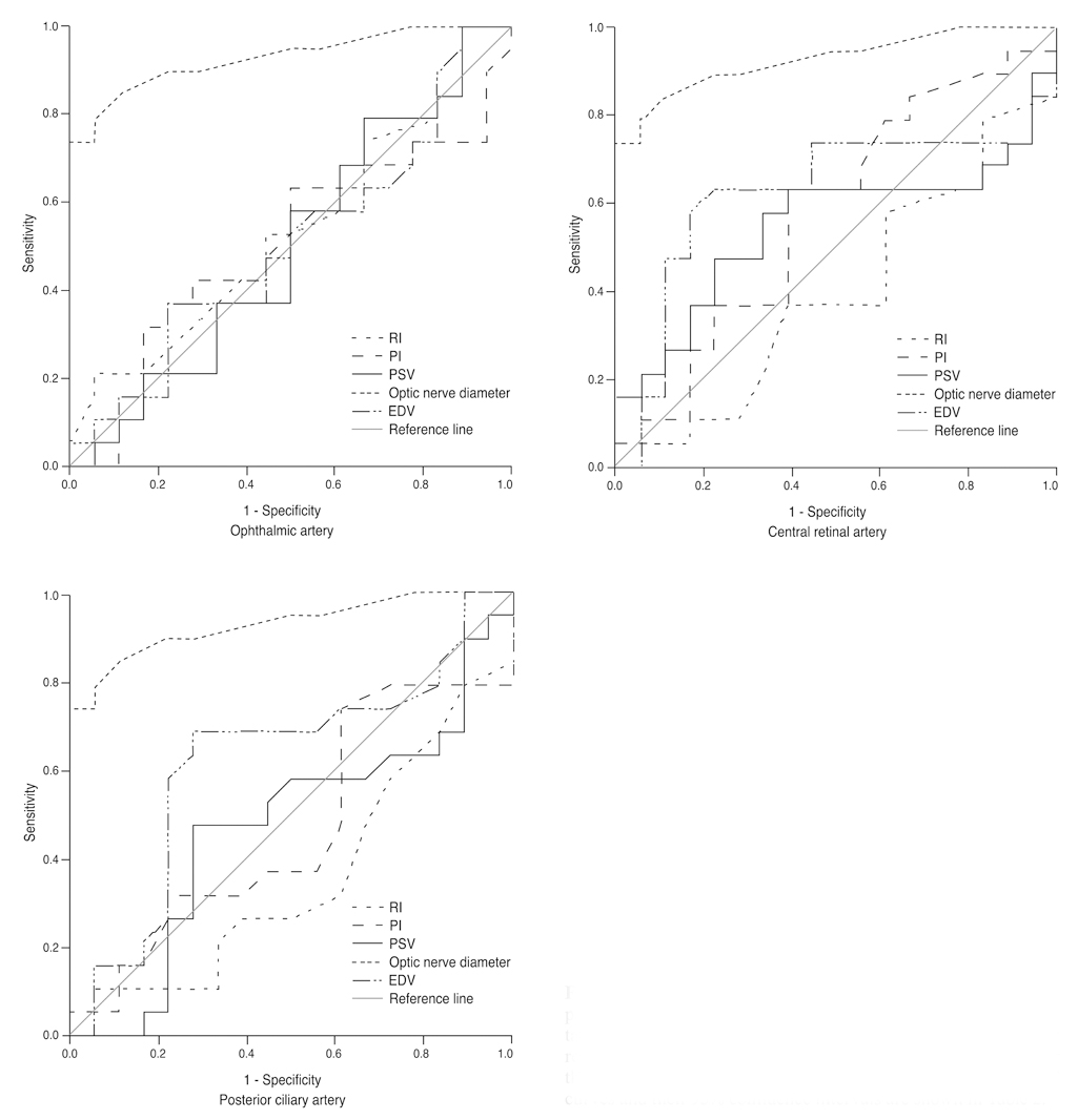
Fig. 1 Receiver operating characteristic (ROC) curves for the peak systolic velocity (PSV), end-diastolic velocity (EDV), resistance index (RI), and pulsatile index (PI) of ophthalmic, central retinal and posterior ciliary arteries and optic nerve diameter for the diagnosis of optic neuritis. The estimated area under the ROC curves and their 95% confidence intervals are shown in Table 2.
Reference
-
1. Acevedo AR, Nava C, Arriada N, et al. Cardiovascular dysfunction in multiple sclerosis. Acta Neurol Scand. 2000. 101:85–88.2. Flachenecker P, Wolf A, Krauser M, et al. Cardiovascular autonomic dysfunction in multiple sclerosis: correlation with orthostatic intolerance. J Neurol. 1999. 246:578–586.3. Speciale L, Sarasella M, Ruzzante S, et al. Endothelin and nitric oxide levels in cerebrospinal fluid of patients with multiple sclerosis. J Neurovirol. 2000. 6:Suppl 2. S62–S66.4. Elvin A, Andersson T, Soderstrom M. Optic neuritis: Doppler ultrasonography compared with MR and correlated with visual evoked potential assessments. Acta Radiol. 1998. 39:243–248.5. Karaali K, Senol U, Aydin H, et al. Optic neuritis: evaluation with orbital Doppler sonography. Radiology. 2003. 226:355–358.6. Akarsu C, Bilgili MY. Color Doppler imaging in ocular hypertension and open-angle glaucoma. Graefes Arch Clin Exp Ophthalmol. 2004. 242:125–129.7. Hradilek P, Zapletalova O, Dolezil D, Skoloudik D. Acute optic ne uritis in multiple sclerosis: evaluation of hemodynamics in the ophthalmic artery with colour Doppler imaging. Neuro-ophthalmology. 2005. 29:161–164.8. Akarsu C, Tan FU, Kendi T. Color Doppler imaging in optic neuritis with multiple sclerosis. Graefes Arch Clin Exp Ophthalmol. 2004. 242:990–994.9. Pache M, Kaiser HJ, Akhalbedashvili N, et al. Extraocular blood flow and endothelin-1 plasma levels in patients with multiple sclerosis. Eur Neurol. 2003. 49:164–168.10. Goh KY, Kay MD, Hughes JR. Orbital color Doppler imaging in nonischemic optic atrophy. Ophthalmology. 1997. 104:330–333.11. Hradilek P, Stourac P, Bar M, et al. Colour Doppler imaging evaluation of blood flow parameters in the ophthalmic artery in acute and chronic phases of optic neuritis in multiple sclerosis. Acta Ophthalmol. 2009. 87:65–70.12. Modrzejewska M, Karczewicz D, Wilk G. Assessment of blood flow velocity in eyeball arteries in multiple sclerosis patients with past retrobulbar optic neuritis in color Doppler ultrasonography. Klin Oczna. 2007. 109:183–186.13. Erdogmus S, Govsa F. Topography of the posterior arteries supplying the eye and relations to the optic nerve. Acta Ophthalmol Scand. 2006. 84:642–649.14. Baxter GM, Williamson TH. Color Doppler imaging of the eye: normal ranges, reproducibility, and observer variation. J Ultrasound Med. 1995. 14:91–96.15. Dees C, Buimer R, Dick AD, Atta HR. Ultrasonographic investigation of optic neuritis. Eye (Lond). 1995. 9(Pt 4):488–494.16. Williamson TH, Harris A. Color Doppler ultrasound imaging of the eye and orbit. Surv Ophthalmol. 1996. 40:255–267.17. Planiol T, Pourcelot L, Itti R. The carotid and cerebral circulations. Advances in its study by external physical methods. Principles, normal recordings, adopted parameters. Nouv Presse Med. 1973. 2:2451–2456.18. Gosling RG, King DH. Arterial assessment by Doppler-shift ultrasound. Proc R Soc Med. 1974. 67(6 Pt 1):447–449.19. DeLong ER, DeLong DM, Clarke-Pearson DL. Comparing the areas under two or more correlated receiver operating characteristic curves: a nonparametric approach. Biometrics. 1988. 44:837–845.20. Gerling J, Janknecht P, Hansen LL, Kommerell G. Diameter of the optic nerve in idiopathic optic neuritis and in anterior ischemic optic neuropathy. Int Ophthalmol. 1997. 21:131–135.21. McLeod D, Marshall J, Kohner EM. Role of axoplasmic transport in the pathophysiology of ischaemic disc swelling. Br J Ophthalmol. 1980. 64:247–261.22. Mostbeck GH, Gossinger HD, Mallek R, et al. Effect of heart rate on Doppler measurements of resistive index in renal arteries. Radiology. 1990. 175:511–513.23. Guthoff RF, Berger RW, Winkler P, et al. Doppler ultrasonography of the ophthalmic and central retinal vessels. Arch Ophthalmol. 1991. 109:532–536.24. MacKenzie F, De Vermette R, Nimrod C, et al. Doppler sonographic studies on the ophthalmic and central retinal arteries in the gravid woman. J Ultrasound Med. 1995. 14:643–647.
