Digital Tomosynthesis for PNS Evaluation: Comparisons of Patient Exposure and Image Quality with Plain Radiography
- Affiliations
-
- 1Department of Radiology, Samsung Medical Center, Sungkyunkwan University School of Medicine, Seoul 135-710, Korea. mjchung@skku.edu
- 2Department of Radiology, Bundang Jesaeng General Hospital, Sungnam 463-774, Korea.
- 3Division of Pulmonology, Department of Internal Medicine, Samsung Medical Center, Sungkyunkwan University School of Medicine, Seoul 135-710, Korea.
- KMID: 1245377
- DOI: http://doi.org/10.3348/kjr.2012.13.2.136
Abstract
OBJECTIVE
We investigated low dose digital tomosynthesis (DT) for the evaluation of the paranasal sinus (PNS), and compared its diagnostic accuracy with a PNS radiography series (XR).
MATERIALS AND METHODS
We enrolled 43 patients for whom XR, PNS DT, and OMU CT were performed. We measured effective doses (EDs) of XR, DT, and OMU CT using Monte Carlo simulation software. Two radiologists performed independent observation of both XR and DT. For seven PNSs, they scored anatomic conspicuity of sinuses and confidence on the presence of sinusitis using nine point scales. OMU CT was observed by the third radiologist and the findings were regarded as reference standard. We compared scores for conspicuity and sinusitis confidence between XR and DT.
RESULTS
Mean EDs were 29 +/- 6 microSv, 48 +/- 10 microSv, and 980 +/- 250 microSv, respectively, for XR, DT, and CT. Mean scores for conspicuity were 6.3 and 7.4, respectively, for XR and DT. Sensitivity per patient basis for sinusitis detection were 52% and 96%, respectively, for XR and DT in observer 1 (p = 0.001) and 80% and 92% for observer 2 (p = 0.25). Specificities for sinusitis exclusion were 100% for both XR and DT for observer 1 and 89% and 100% for observer 2 (p = 0.50). Accuracies for sinusitis diagnosis were 72% and 98%, respectively, for XR and DT for observer 1 (p = 0.001) and 84% and 95% for observer 2 (p = 0.125).
CONCLUSION
Patient radiation dose from low dose DT is comparable with that of PNS XR. Diagnostic sensitivity of DT for sinusitis was superior to PNS XR.
MeSH Terms
Figure
-
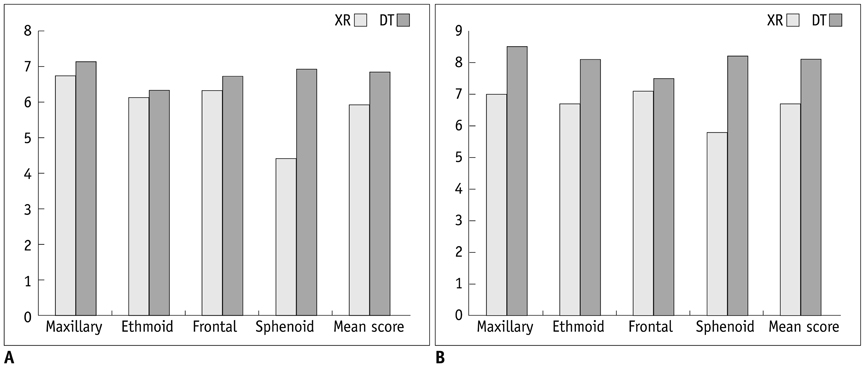
Fig. 1 Scores for anatomic conspicuity for radiography (XR) and digital tomosynthesis (DT) for demarcation of paranasal sinuses. A. For observer 1, mean socres are 5.9 for XR and 6.8 for DT (p < 0.01). B. For observer 2, mean socres are 6.7 for XR and 8.1 for DT (p < 0.01).
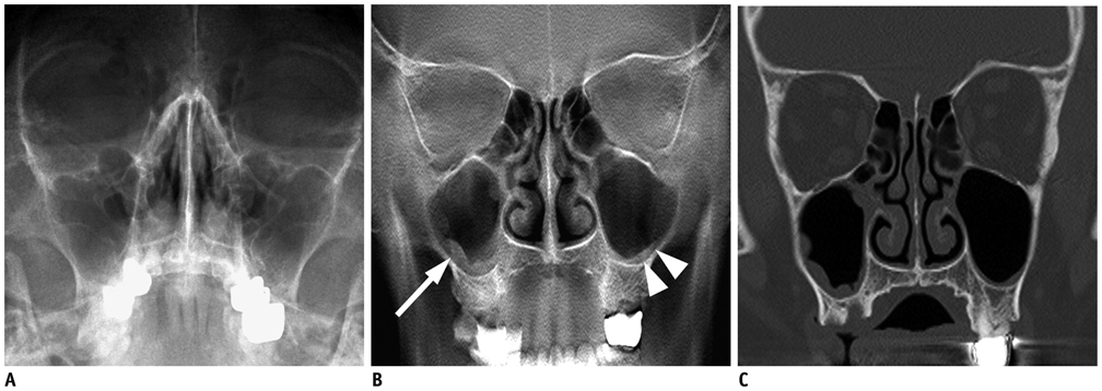
Fig. 2 Images from 65-year-old man with fever. A. Water's view radiograph shows subtle periosteal blurring of right maxillary sinuses. Both observers missed presence of sinusitis. B. Tomosynthesis image shows lobulating mucoperiosteal thickening in right mixillary sinus (arrow) and minimal and smooth thickening in left maxillary sinus (arrowheads). C. CT image confirms presence of mucoperiosteal thickening in both maxillary sinuses.
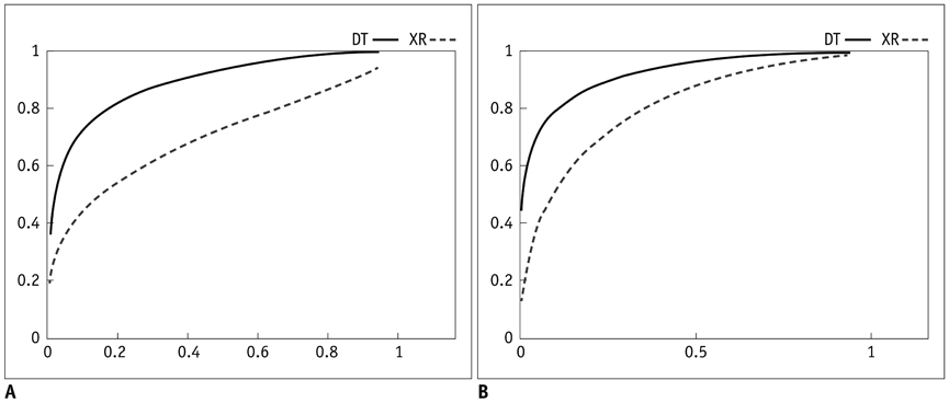
Fig. 3 Receiver operator characteristic curve (ROC curve) from radiography (XR) and digital tomosynthesis (DT) for detection of paranasal sinusitis. A. For observer 1, areas under curve (Az) are 0.700 and 0.891, respectively, for XR and DT (p < 0.05). B. For observer 2, Azs are 0.813 and 0.924, respectively, for XR and DT (p < 0.05).
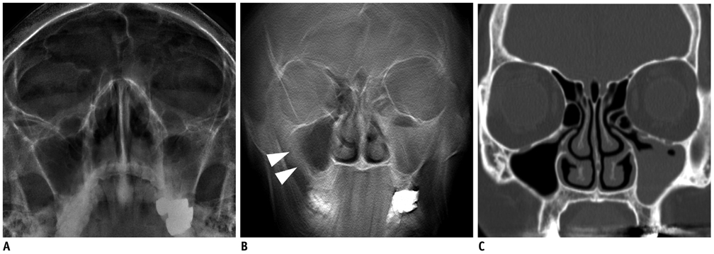
Fig. 4 Images from 68-year-old woman with cough. A. Water's view radiograph shows subtle fluid filled left maxillary sinus. There is no motion artifact. B. Tomosynthesis image shows image blurring in right maxillary sinus (arrowheads) which mimic mucoperiosteal thickening. Note prominent motion artifact in mandible area. C. CT image confirms presence of left maxillary sinusitis. Note that right maxillary sinus is normal.
Cited by 1 articles
-
Assessment of Maxillary Sinus Wall Thickness with Paranasal Sinus Digital Tomosynthesis and CT
Jieun Byun, Sung Shine Shim, Yookyung Kim, Kyoung Ae Kong
J Korean Soc Radiol. 2017;76(5):314-321. doi: 10.3348/jksr.2017.76.5.314.
Reference
-
1. Dobbins JT 3rd, Godfrey DJ. Digital x-ray tomosynthesis: current state of the art and clinical potential. Phys Med Biol. 2003. 48:R65–R106.2. Grant DG. Tomosynthesis: a three-dimensional radiographic imaging technique. IEEE Trans Biomed Eng. 1972. 19:20–28.3. Johnsson AA, Vikgren J, Svalkvist A, Zachrisson S, Flinck A, Boijsen M, et al. Overview of two years of clinical experience of chest tomosynthesis at Sahlgrenska University Hospital. Radiat Prot Dosimetry. 2010. 139:124–129.4. Dobbins JT 3rd, McAdams HP. Chest tomosynthesis: technical principles and clinical update. Eur J Radiol. 2009. 72:244–251.5. Dobbins JT 3rd, McAdams HP, Godfrey DJ, Li CM. Digital tomosynthesis of the chest. J Thorac Imaging. 2008. 23:86–92.6. Vikgren J, Zachrisson S, Svalkvist A, Johnsson AA, Boijsen M, Flinck A, et al. Comparison of chest tomosynthesis and chest radiography for detection of pulmonary nodules: human observer study of clinical cases. Radiology. 2008. 249:1034–1041.7. James TD, McAdams HP, Song JW, Li CM, Godfrey DJ, DeLong DM, et al. Digital tomosynthesis of the chest for lung nodule detection: interim sensitivity results from an ongoing NIH-sponsored trial. Med Phys. 2008. 35:2554–2557.8. Sharifian H, Sharif Kashani S, Mazaher H, Abhari M. Assessment of the accuracy of paranasal sinuses limited CT scans in the diagnosis of Sinusitis. Iran J Radiol. 2006. 3:225–228.9. Leibovitch I, Goldberg RA, Selva D. Paranasal sinus inflammation and non-specific orbital inflammatory syndrome: an uncommon association. Graefes Arch Clin Exp Ophthalmol. 2006. 244:1391–1397.10. Roberts DN, Hampal S, East CA, Lloyd GA. The diagnosis of inflammatory sinonasal disease. J Laryngol Otol. 1995. 109:27–30.11. Robinson KE. Roentgenographic manifestations of benign paranasal disease. Ear Nose Throat J. 1984. 63:144–149.12. Dolan KD. Paranasal sinus radiology, Part 3A: sphenoidal sinus. Head Neck Surg. 1982. 5:164–176.13. White PS, Cowan IA, Robertson MS. Limited CT scanning techniques of the paranasal sinuses. J Laryngol Otol. 1991. 105:20–23.14. Wippold FJ 2nd, Levitt RG, Evens RG, Korenblat PE, Hodges FJ 3rd, Jost RG. Limited coronal CT: an alternative screening examination for sinonasal inflammatory disease. Allergy Proc. 1995. 16:165–169.15. White PS, Robinson JM, Stewart IA, Doyle T. Computerized tomography mini-series: an alternative to standard paranasal sinus radiographs. Aust N Z J Surg. 1990. 60:25–29.16. Sabol JM. A Monte Carlo estimation of effective dose in chest tomosynthesis. Med Phys. 2009. 36:5480–5487.17. Huda W, Ogden KM, Khorasani MR. Converting dose-length product to effective dose at CT. Radiology. 2008. 248:995–1003.18. Baldwin P. Digital breast tomosynthesis. Radiol Technol. 2009. 81:57M–74M.19. Gennaro G, Toledano A, di Maggio C, Baldan E, Bezzon E, La Grassa M, et al. Digital breast tomosynthesis versus digital mammography: a clinical performance study. Eur Radiol. 2010. 20:1545–1553.20. Michell M, Wasan R, Whelehan P, Iqbal A, Lawinski C, Donaldson A, et al. Digital breast tomosynthesis: a comparison of the accuracy of digital breast tomosynthesis, two-dimensional digital mammography and two-dimensional screening mammography (film-screen). Breast Cancer Res. 2009. 11:Suppl 2. O1.21. Gur D, Abrams GS, Chough DM, Ganott MA, Hakim CM, Perrin RL, et al. Digital breast tomosynthesis: observer performance study. AJR Am J Roentgenol. 2009. 193:586–591.22. Schulz-Wendtland R, Fuchsjäger M, Wacker T, Hermann KP. Digital mammography: an update. Eur J Radiol. 2009. 72:258–265.23. Duryea J, Dobbins JT 3rd, Lynch JA. Digital tomosynthesis of hand joints for arthritis assessment. Med Phys. 2003. 30:325–333.24. Zachrisson S, Vikgren J, Svalkvist A, Johnsson AA, Boijsen M, Flinck A, et al. Effect of clinical experience of chest tomosynthesis on detection of pulmonary nodules. Acta Radiol. 2009. 50:884–891.25. Tingberg A. X-ray tomosynthesis: a review of its use for breast and chest imaging. Radiat Prot Dosimetry. 2010. 139:100–107.26. Lewin JM, Niklason L. Advanced applications of digital mammography: tomosynthesis and contrast-enhanced digital mammography. Semin Roentgenol. 2007. 42:243–252.27. Bakic PR, Carton AK, Kontos D, Zhang C, Troxel AB, Maidment AD. Breast percent density: estimation on digital mammograms and central tomosynthesis projections. Radiology. 2009. 252:40–49.28. Teertstra HJ, Loo CE, van den Bosch MA, van Tinteren H, Rutgers EJ, Muller SH, et al. Breast tomosynthesis in clinical practice: initial results. Eur Radiol. 2010. 20:16–24.29. Zhang J, Wu QJ, Godfrey DJ, Fatunase T, Marks LB, Yin FF. Comparing digital tomosynthesis to cone-beam CT for position verification in patients undergoing partial breast irradiation. Int J Radiat Oncol Biol Phys. 2009. 73:952–957.30. Kim EY, Chung MJ, Lee HY, Koh WJ, Jung HN, Lee KS. Pulmonary mycobacterial disease: diagnostic performance of low-dose digital tomosynthesis as compared with chest radiography. Radiology. 2010. 257:269–277.31. Dobbins JT 3rd. Tomosynthesis imaging: at a translational crossroads. Med Phys. 2009. 36:1956–1967.32. Taourel P, Merigeaud S, Aubert E, Millet I, Curros Doyon F, Lacroix J, et al. [Tomosynthesis: luxury or necessity?]. J Radiol. 2009. 90:1813–1821.
- Full Text Links
-
- Actions
-
Cited
- CITED
-
- Close
- Share
-
- Similar articles
-
- Usefulness of Digital Tomosynthesis for the Detection of Airway Obstruction: A Case Report of Bronchial Carcinosarcoma
- Objective and Quantitative Evaluation of Image Quality Using Fuzzy Integral: Phantom Study
- A Comparative Study of Image Quality and Radiation Dose with Changes in Tube Voltage and Current for a Digital Chest Radiography
- Digital Tomosynthesis of the Chest: Comparison of Patient Exposure Dose and Image Quality between Standard Default Setting and Low Dose Setting
- Compressed-sensing (CS)-based Image Deblurring Scheme with a Total Variation Regularization Penalty for Improving Image Characteristics in Digital Tomosynthesis (DTS)
