A Case of Hypopharyngeal Cancer Treated by Endoscopic Submucosal Dissection
- Affiliations
-
- 1Division of Gastroenterology, Department of Internal Medicine, Guro Hospital, Korea University College of Medicine, Seoul, Korea. gi7pjj@yahoo.co.kr
- 2Department of Pathology, Guro Hospital, Korea University College of Medicine, Seoul, Korea.
- KMID: 1245209
- DOI: http://doi.org/10.4166/kjg.2012.59.3.239
Abstract
- Recent advances in endoscopic instruments, including narrow-band imaging (NBI) and magnification endoscopy, allowed dramatic increase in the early diagnosis of hypopharyngeal cancers. In addition, endoscopic mucosal resection or endoscopic submucosal dissection has recently been used for the treatment of hypopharyngeal cancer at an early stage, especially in Japan. However, to date, there is no published report in Korea. A 68-year-old man was admitted for preoperative evaluation and treatment for known esophageal cancer initially diagnosed at a local clinic. During the evaluation, magnifying endoscopy combined with the NBI system revealed a concurrent hypopharyngeal cancer not detected by initial conventional endoscopy. In this case report, we describe for the first time in Korea a case of early stage hypopharyngeal carcinoma that was successfully treated by endoscopic submucosal dissection with a review of literature.
MeSH Terms
Figure
-

Fig. 1 Conventional (A) and magnifying (B) endoscopy combined with the narrow band imaging system showing a clearly demarcated brownish colored area, and increased numbers of distorted intraepithelial papillary capillary at the left pyriform sinus, respectively.
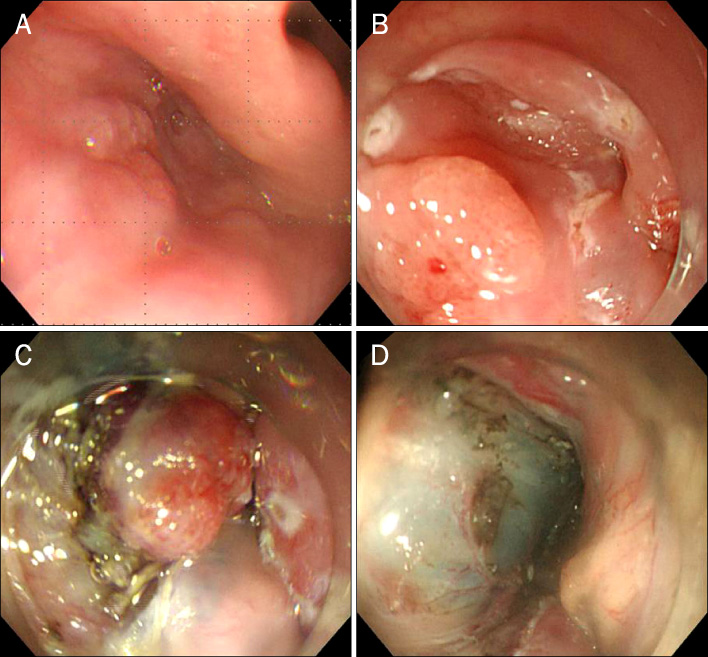
Fig. 2 Serial endoscopic images of endoscopic submucosal dissection procedure for the removal of an early hypopharyngeal cancer showing a 10×8 mm sized, slightly elevated lesion with surface irregularity and hyperemia on the left pyriform sinus (A), markings around the lesion with a needle knife (B), circumferential mucosal incision and submucosal dissection with an hook and IT knife (C), and artificial ulcer after complete en-bloc resection (D).
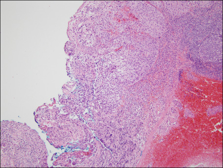
Fig. 3 Photomicrograph of the endoscopic submucosal dissection specimen. The tumor was moderately differentiated squamous cell carcinoma limited to the subepithelial layer showing squamous differentiation with distinct nuclear pleomorphism, mitotic activity and rare keratinization limited to the subepithelial layer (H&E, ×100).

Fig. 4 Follow-up endoscopic image. 2 months after endoscopic submucosal dissection (ESD), post ESD scar with a tiny erosion on left hypopharynx.
Reference
-
1. Triboulet JP, Mariette C, Chevalier D, Amrouni H. Surgical management of carcinoma of the hypopharynx and cervical esophagus: analysis of 209 cases. Arch Surg. 2001. 136:1164–1170.2. Pesko P, Sabljak P, Bjelovic M, et al. Surgical treatment and clinical course of patients with hypopharyngeal carcinoma. Dis Esophagus. 2006. 19:248–253.3. de Graeff A, de Leeuw JR, Ros WJ, Hordijk GJ, Blijham GH, Winnubst JA. Long-term quality of life of patients with head and neck cancer. Laryngoscope. 2000. 110:98–106.4. Shimizu Y, Tsukagoshi H, Fujita M, et al. Head and neck cancer arising after endoscopic mucosal resection for squamous cell carcinoma of the esophagus. Endoscopy. 2003. 35:322–326.5. Muto M, Nakane M, Katada C, et al. Squamous cell carcinoma in situ at oropharyngeal and hypopharyngeal mucosal sites. Cancer. 2004. 101:1375–1381.6. Lehman G, Compton M, Meadows J, Elmore M. Screening examination of the larynx and pharynx during upper gastrointestinal panendoscopy. Gastrointest Endosc. 1982. 28:176–178.7. Watanabe S, Matsuda K, Arima K, et al. Detection of subclinical disorders of the hypopharynx and larynx by gastrointestinal endoscopy. Endoscopy. 1996. 28:295–298.8. Slaughter DP, Southwick HW, Smejkal W. Field cancerization in oral stratified squamous epithelium; clinical implications of multicentric origin. Cancer. 1953. 6:963–968.9. Muto M, Katada C, Sano Y, Yoshida S. Narrow band imaging: a new diagnostic approach to visualize angiogenesis in superficial neoplasia. Clin Gastroenterol Hepatol. 2005. 3:7 Suppl 1. S16–S20.10. Takenaka R, Kawahara Y, Okada H, et al. Narrow-band imaging provides reliable screening for esophageal malignancy in patients with head and neck cancers. Am J Gastroenterol. 2009. 104:2942–2948.11. Evrard S, Le Moine O, Hassid S, Devière J. Zenker's diverticulum: a new endoscopic treatment with a soft diverticuloscope. Gastrointest Endosc. 2003. 58:116–120.12. Lee BJ, Park JJ, Joo MK, et al. The feasibility and safety of endoscopic resection for benign hypopharyngeal tumors. Hepatogastroenterology. 2009. 56:636–640.13. Shimizu Y, Yamamoto J, Kato M, et al. Endoscopic submucosal dissection for treatment of early stage hypopharyngeal carcinoma. Gastrointest Endosc. 2006. 64:255–259.14. Iizuka T, Kikuchi D, Hoteya S, Yahagi N, Takeda H. Endoscopic submucosal dissection for treatment of mesopharyngeal and hypopharyngeal carcinomas. Endoscopy. 2009. 41:113–117.15. Yoshida T, Shimizu Y, Hirota J, et al. Early-stage laryngeal squamous cell carcinoma of the epiglottis treated by endoscopic submucosal dissection. Endoscopy. 2008. 40:Suppl 2. E204–E205.16. Shimizu Y, Yoshida T, Kato M, et al. Long-term outcome after endoscopic resection in patients with hypopharyngeal carcinoma invading the subepithelium: a case series. Endoscopy. 2009. 41:374–376.17. Suzuki H, Saito Y, Oda I, Nonaka S, Nakanishi Y. Feasibility of endoscopic mucosal resection for superficial pharyngeal cancer: a minimally invasive treatment. Endoscopy. 2010. 42:1–7.18. Iizuka T, Kikuchi D, Hoteya S, et al. Clinical advantage of endoscopic submucosal dissection over endoscopic mucosal resection for early mesopharyngeal and hypopharyngeal cancers. Endoscopy. 2011. 43:839–843.
- Full Text Links
-
- Actions
-
Cited
- CITED
-
- Close
- Share
-
- Similar articles
-
- History and Development of Accessories for Endoscopic Submucosal Dissection
- The Clinical Accuracy of Endoscopic Ultrasonography and White Light Imaging in Gastric Endoscopic Submucosal Dissection
- A Case of Pneumorrhachis and Pneumoscrotum Following Colon Endoscopic Submucosal Dissection
- Future Development of Endoscopic Accessories for Endoscopic Submucosal Dissection
- Pathological Interpretation of Gastric Tumors in Endoscopic Submucosal Dissection
