Plexiform Angiomyxoid Myofibroblastic Tumor of the Stomach: A Case Report
- Affiliations
-
- 1Department of Pathology, Yeungnam University College of Medicine, Daegu, Korea. ykbae@ynu.ac.kr
- KMID: 1123453
- DOI: http://doi.org/10.3346/jkms.2011.26.11.1508
Abstract
- Plexiform angiomyxoid myofibroblastic tumor (PAMT) is a recently described mesenchymal tumor of the stomach. We report the first case of PAMT in Korea. A 52-yr-old man underwent esophagogastroduodenoscopy due to dyspepsia for 2 yr. There was a submucosal mass with small mucosal ulceration in the gastric antrum. The tumor measured 3.5 x 2.3 cm in size and showed multinodular plexiform growth pattern of bland-looking spindle cells separated by an abundant myxoid or fibromyxoid matrix rich in small thin-walled blood vessels. The tumor cells were negative for CD117 (c-KIT), CD34 and S-100 protein, but diffusely positive for smooth muscle actin consistent with predominant myofibroblastic differentiation. The patient is doing well without recurrence or metastasis for 5 months after surgery. Although there have been limited follow-up data, PAMT is regarded as a benign gastric neoplasm with histological and immunohistochemical charateristics distinguished from gastrointestinal stromal tumor and other mesenchymal tumors of the stomach.
MeSH Terms
Figure
-

Fig. 1 Endoscopic examination of the tumor. (A) The stomach shows a submucosal mass with small mucosal ulceration in the antrum. (B) The biopsied mucosa shows an ill-defined myxoid lesion with loosely scattered spindle cells (H&E, × 40). The spindle tumor cells were positive for smooth muscle actin (C) and negative for CD117 (D) immunostains (× 200).
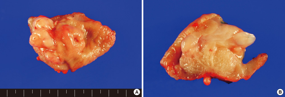
Fig. 2 Gross examination of the excised specimen. (A) There is a protruding multinodular glistening mass, mainly located in the submucosa. (B) Cut surface showed myxoid nature and unclear margin of the tumor.

Fig. 3 Microscopic examination of the tumor. (A) The tumor showed plexiform growth pattern dissecting the muscularis propria (H&E, × 10). The bland-looking spindle tumor cells were separated by abundant myxoid (B) or fibromyxoid stroma (C) (H&E, × 100). (D) Palisading arrangement of tumor cells was noted (H&E, × 200). (E) There were numerous thin-walled vessels in the myxoid stroma (H&E, × 200). (F) The tumor cells were focally positive for desmin immunostain (× 200).
Reference
-
1. Miettinen M, Fletcher CD, Kindblom LG, Tsui WM. Bosman FT, Carneiro F, Hruban R, Theise ND, editors. Mesenchymal tumours of the stomach. WHO classification of tumours of the digestive system. 2010. Lyon: IARC;74–79.2. Takahashi Y, Shimizu S, Ishida T, Aita K, Toida S, Fukusato T, Mori S. Plexiform angiomyxoid myofibroblastic tumor of the stomach. Am J Surg Pathol. 2007. 31:724–728.3. Miettinen M, Makhlouf HR, Sobin LH, Lasota J. Plexiform fibromyxoma: a distinctive benign gastric antral neoplasm not to be confused with a myxoid GIST. Am J Surg Pathol. 2009. 33:1624–1632.4. Rau TT, Hartmann A, Dietmaier W, Schmitz J, Hohenberger W, Hofstaedter F, Katenkamp K. Plexiform angiomyxoid myofibroblastic tumour: differential diagnosis of gastrointestinal stromal tumour in the stomach. J Clin Pathol. 2008. 61:1136–1137.5. Galant C, Rousseau E, Ho Minh Duc DK, Pauwels P. Plexiform angiomyxoid myofibroblastic tumor of the stomach. Am J Surg Pathol. 2008. 32:1910.6. Yoshida A, Klimstra DS, Antonescu CR. Plexiform angiomyxoid tumor of the stomach. Am J Surg Pathol. 2008. 32:1910–1912.7. Pailoor J, Mun KS, Chen CT, Pillay B. Plexiform angiomyxoid myofibroblastic tumour of the stomach. Pathology. 2009. 41:698–699.8. Takahashi Y, Suzuki M, Fukusato T. Plexiform angiomyxoid myofibroblastic tumor of the stomach. World J Gastroenterol. 2010. 16:2835–2840.9. Sing Y, Subayan S, Mqadi B, Ramdial PK, Reddy J, Moodley MS, Bux S. Gastric plexiform angiomyxoid myofibroblastic tumor. Pathol Int. 2010. 60:621–625.10. Tan CY, Santos LD, Biankin A. Plexiform angiomyxoid myofibroblastic tumor of the stomach: a case report. Pathology. 2010. 42:581–583.
- Full Text Links
-
- Actions
-
Cited
- CITED
-
- Close
- Share
-
- Similar articles
-
- Plexiform Angiomyxoid Myofibroblastic Tumor of the Stomach: Report of a Case and Review of the Literature
- Plexiform Angiomyxoid Myofibroblastic Tumor of the Stomach: Report of Two Cases and Review of the Literature
- Plexiform Angiomyxoid Myofibroblastic Tumor of the Stomach: a Rare Case
- Inflammatory Myofibroblastic Tumor of Nasal Septum after Septoplasty: A Case Report
- A Case of Myxoid Plexiform Fibrohistiocytic Tumor
