Redistributed Regional Ventilation after the Administration of a Bronchodilator Demonstrated on Xenon-Inhaled Dual-Energy CT in a Patient with Asthma
- Affiliations
-
- 1Department of Radiology and the Research Institute of Radiology, Asan Medical Center, University of Ulsan College of Medicine, Seoul 138-736, Korea. hwgoo@amc.seoul.kr
- 2Department of Pediatrics, Asan Medical Center, University of Ulsan College of Medicine, Seoul 138-736, Korea.
- KMID: 1122337
- DOI: http://doi.org/10.3348/kjr.2011.12.3.386
Abstract
- We report here on the redistributed regional ventilation abnormalities after the administration of a bronchodilator and as seen on xenon-inhaled dual-energy CT in a patient with asthma. The improved ventilation seen in the right lower lobe and the decreased ventilation seen in the right middle lobe after the administration of a bronchodilator on xenon-inhaled dual-energy CT could explain a positive bronchodilator response on a pulmonary function test. These changes may reflect the heterogeneity of the airway responsiveness to a bronchodilator in patients with asthma.
MeSH Terms
Figure
-
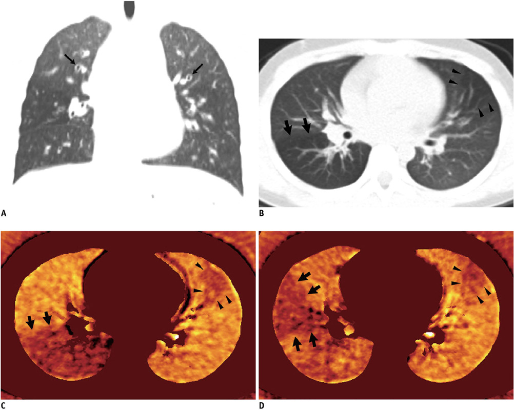
Fig. 1 7-year-old girl with bronchial asthma. A. Coronal weighted-average CT image after administration of bronchodilator shows wall thickenings of central airways (arrows). Hyperlucency that suggests air trapping is noted in right middle lobe. B. Axial weighted-average CT image before administration of bronchodilator shows focal hyperlucent areas in superior segment of right lower lobe (arrows) and in left upper lobe lingular division (arrowheads). C. Axial xenon map before administration of bronchodilator reveals severe ventilation defect in superior segment of right lower lobe (arrows) and milder subsegmental ventilation defect in left upper lobe lingular division (arrowheads). D. Axial xenon map after administration of bronchodilator demonstrates redistributed regional ventilation in right lung. Xenon values in superior segment of right lower lobe are remarkably increased, while those in right middle lobe and especially those in lateral segment (arrows) are decreased. On other hand, subsegmental defect in left upper lobe lingular division (arrowheads) appears unchanged. Of note, xenon values in normally ventilated lung regions appear slightly lower than those on xenon map before administration of bronchodilator (C) and this was probably due to slight difference in inhaled xenon concentration and respiration level between two dual-energy CT scans.
Cited by 1 articles
-
Dual-Energy CT: New Horizon in Medical Imaging
Hyun Woo Goo, Jin Mo Goo
Korean J Radiol. 2017;18(4):555-569. doi: 10.3348/kjr.2017.18.4.555.
Reference
-
1. Chae EJ, Seo JB, Goo HW, Kim N, Song KS, Lee SD, et al. Xenon ventilation CT with a dual-energy technique of dual-source CT: initial experience. Radiology. 2008. 248:615–624.2. Goo HW, Chae EJ, Seo JB, Hong SJ. Xenon ventilation CT using a dual-source dual-energy technique: dynamic ventilation abnormality in a child with bronchial atresia. Pediatr Radiol. 2008. 38:1113–1116.3. Chae EJ, Seo JB, Kim N, Song KS, Shin JH, Kim TH, et al. Collateral ventilation in a canine model with bronchial obstruction: assessment with xenon-enhanced dual-energy CT. Radiology. 2010. 255:790–798.4. Goo HW, Yang DH, Hong SJ, Yu J, Kim BJ, Seo JB, et al. Xenon ventilation CT using dual-source and dual-energy technique in children with bronchiolitis obliterans: correlation of xenon and CT density values with pulmonary function test results. Pediatr Radiol. 2010. 40:1490–1497.5. Kang MJ, Park CM, Lee CH, Goo JM, Lee HJ. Dual-energy CT: clinical applications in various pulmonary diseases. Radiographics. 2010. 30:685–698.6. Chae EJ, Seo JB, Lee J, Kim N, Goo HW, Lee HJ, et al. Xenon ventilation imaging using dual-energy computed tomography in asthmatics: initial experience. Invest Radiol. 2010. 45:354–361.7. de Lange EE, Altes TA, Patrie JT, Battiston JJ, Juersivich AP, Mugler JP 3rd, et al. Changes in regional airflow obstruction over time in the lungs of patients with asthma: evaluation with 3He MR imaging. Radiology. 2009. 250:567–575.8. Tzeng YS, Hoffman E, Cook-Granroth J, Gereige J, Mansour J, Washko G, et al. Investigation of hyperpolarized 3He magnetic resonance imaging utility in examining human airway diameter behavior in asthma through comparison with high-resolution computed tomography. Acad Radiol. 2008. 15:799–808.
- Full Text Links
-
- Actions
-
Cited
- CITED
-
- Close
- Share
-
- Similar articles
-
- Collateral Ventilation to Congenital Hyperlucent Lung Lesions Assessed on Xenon-Enhanced Dynamic Dual-Energy CT: an Initial Experience
- Visual and Quantitative Assessments of Regional Xenon-Ventilation Using Dual-Energy CT in Asthma-ChronicObstructive Pulmonary Disease Overlap Syndrome:A Comparison with Chronic Obstructive PulmonaryDisease
- Collateral Ventilation Quantification Using Xenon-Enhanced Dynamic Dual-Energy CT: Differences between Canine and Swine Models of Bronchial Occlusion
- Measurement and Imaging of Regional Cerebral Blood Flow(rCBF) with Xenon-Enhanced CT(XECT)
- Dual-Energy CT: New Horizon in Medical Imaging
