Korean J Ophthalmol.
2012 Feb;26(1):6-9. 10.3341/kjo.2012.26.1.6.
Posterior Corneal Curvature Assessment after Epi-LASIK for Myopia: Comparison of Orbscan II and Pentacam Imaging
- Affiliations
-
- 1Department of Ophthalmology and Visual Science, Seoul St. Mary's Hospital, The Catholic University of Korea College of Medicine, Seoul, Korea. ckjoo@catholic.ac.kr
- 2Catholic Institute for Visual Science, The Catholic University of Korea College of Medicine, Seoul, Korea.
- KMID: 1120174
- DOI: http://doi.org/10.3341/kjo.2012.26.1.6
Abstract
- PURPOSE
To compare the changes in posterior corneal curvature using scanning slit topography (Orbscan II) and Scheimpflug imaging (Pentacam) before and after Epi-laser in situ keratomileusis (LASIK) for myopia.
METHODS
In a prospective observational case-series study, 20 myopic patients having undergone Epi-LASIK were examined serially with two different devices, Orbscan II and Pentacam, preoperatively and one month postoperatively. Posterior central elevation (PCE) and posterior maximal elevation (PME) were compared between the two devices, and the changes in parameters after Epi-LASIK were analyzed using a difference map.
RESULTS
All parameters (preoperative and postoperative PCE and preoperative and postoperative PME) that were measured using the Orbscan II were significantly greater compared to those of the Pentacam (for all p < 0.001). PCE and PME were significantly increased one month postoperatively in the Orbscan II measurements (p < 0.05) but were not significantly increased in the Pentacam measurements. Also, DeltaPCE and DeltaPME, in the difference map obtained by each serial scanning, were significantly greater in the Orbscan II measurements than with the Pentacam (p = 0.012, p = 0.016).
CONCLUSIONS
The Pentacam measurements displayed significantly reduced values in all parameters related to posterior corneal elevation compared to those of the Orbscan II. The Pentacam showed no significant change in posterior corneal curvature after Epi-LASIK, based on the difference map.
Keyword
MeSH Terms
Figure
-
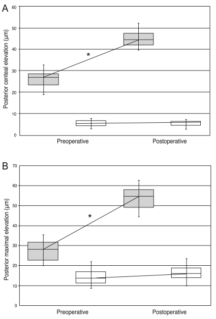
Fig. 1 Intra-device comparison of pre-operative and post-operative values (*p < 0.05). PCE = posterior central elevation; PME = posterior maximal elevation.
Reference
-
1. Wang Z, Chen J, Yang B. Posterior corneal surface topographic changes after laser in situ keratomileusis are related to residual corneal bed thickness. Ophthalmology. 1999; 106:406–409. PMID: 9951499.2. Kamiya K, Oshika T, Amano S, et al. Influence of excimer laser photorefractive keratectomy on the posterior corneal surface. J Cataract Refract Surg. 2000; 26:867–871. PMID: 10889433.
Article3. Baek T, Lee K, Kagaya F, et al. Factors affecting the forward shift of posterior corneal surface after laser in situ keratomileusis. Ophthalmology. 2001; 108:317–320. PMID: 11158806.
Article4. Seitz B, Torres F, Langenbucher A, et al. Posterior corneal curvature changes after myopic laser in situ keratomileusis. Ophthalmology. 2001; 108:666–672. PMID: 11297480.
Article5. Hernandez-Quintela E, Samapunphong S, Khan BF, et al. Posterior corneal surface changes after refractive surgery. Ophthalmology. 2001; 108:1415–1422. PMID: 11470692.
Article6. Sharma N, Rani A, Balasubramanya R, et al. Posterior corneal topographic changes after partial flap during laser in situ keratomileusis. Br J Ophthalmol. 2003; 87:160–162. PMID: 12543743.
Article7. Lee DH, Seo S, Jeong KW, et al. Early spatial changes in the posterior corneal surface after laser in situ keratomileusis. J Cataract Refract Surg. 2003; 29:778–784. PMID: 12686248.
Article8. Yoshida T, Miyata K, Tokunaga T, et al. Difference map or single elevation map in the evaluation of corneal forward shift after LASIK. Ophthalmology. 2003; 110:1926–1930. PMID: 14522766.
Article9. Miyata K, Tokunaga T, Nakahara M, et al. Residual bed thickness and corneal forward shift after laser in situ keratomileusis. J Cataract Refract Surg. 2004; 30:1067–1072. PMID: 15130645.
Article10. Twa MD, Roberts C, Mahmoud AM, Chang JS Jr. Response of the posterior corneal surface to laser in situ keratomileusis for myopia. J Cataract Refract Surg. 2005; 31:61–71. PMID: 15721697.
Article11. Grzybowski DM, Roberts CJ, Mahmoud AM, Chang JS Jr. Model for nonectatic increase in posterior corneal elevation after ablative procedures. J Cataract Refract Surg. 2005; 31:72–81. PMID: 15721698.
Article12. Kim HJ, Song IK, Joo CK. Keratectasia after laser in situ keratomileusis: clinicopathologic case report. J Korean Ophthalmol Soc. 2005; 46:910–914.13. Cairns G, Ormonde SE, Gray T, et al. Assessing the accuracy of Orbscan II post-LASIK: apparent keratectasia is paradoxically associated with anterior chamber depth reduction in successful procedures. Clin Experiment Ophthalmol. 2005; 33:147–152. PMID: 15807822.
Article14. Ueda T, Nawa Y, Masuda K, et al. Posterior corneal surface changes after hyperopic laser in situ keratomileusis. J Cataract Refract Surg. 2005; 31:2084–2087. PMID: 16412919.
Article15. Ciolino JB, Belin MW. Changes in the posterior cornea after laser in situ keratomileusis and photorefractive keratectomy. J Cataract Refract Surg. 2006; 32:1426–1431. PMID: 16931251.
Article16. Hashemi H, Mehravaran S. Corneal changes after laser refractive surgery for myopia: comparison of Orbscan II and Pentacam findings. J Cataract Refract Surg. 2007; 33:841–847. PMID: 17466859.
Article17. Cairns G, McGhee CN. Orbscan computerized topography: attributes, applications, and limitations. J Cataract Refract Surg. 2005; 31:205–220. PMID: 15721715.
Article18. Kopacz D, Maciejewicz P, Kecik D. Pentacam: the new way for anterior eye segment imaging and mapping. Klin Oczna. 2005; 107:728–731. PMID: 16619832.19. Ha BJ, Kim SW, Kim SW, et al. Pentacam and Orbscan II measurements of posterior corneal elevation before and after photorefractive keratectomy. J Refract Surg. 2009; 25:290–295. PMID: 19370825.20. Ormonde S, Waterman C, McGhee C. Changes in the posterior corneal surface after LASIK. J Cataract Refract Surg. 2004; 30:533–534. PMID: 15050233.
Article21. Nawa Y, Masuda K, Ueda T, et al. Evaluation of apparent ectasia of the posterior surface of the cornea after keratorefractive surgery. J Cataract Refract Surg. 2005; 31:571–573. PMID: 15811747.
Article22. Maldonado MJ, Nieto JC, Diez-Cuenca M, Pinero DP. Repeatability and reproducibility of posterior corneal curvature measurements by combined scanning-slit and placido-disc topography after LASIK. Ophthalmology. 2006; 113:1918–1926. PMID: 16935339.
Article
- Full Text Links
-
- Actions
-
Cited
- CITED
-
- Close
- Share
-
- Similar articles
-
- Central Corneal Thickness Measured by Four Different Methods in Normal and Post-Femtosecond Laser-Assisted LASIK Eyes
- Comparison of Corneal Thickness and Anterior Chamber Depth Measured With Orbscan, Pentacam, and Ultrasound Pachymetry
- Short Term Clinical Results of Laser Epithelial Keratomileusis and Epi-Laser in Situ Keratomileusis for Moderate and High Myopia
- Comparison of Pentacam with Orbscan
- Relationship between Clinical History Method and Orbscan II for Measuring Corneal Power after LASIK
