A Rare Case of Recurrent Myoid Hamartoma Mimicking Malignancy: Imaging Appearances
- Affiliations
-
- 1Department of Pathology, St. Vincent's Hospital, College of Medicine, The Catholic University of Korea, Gyeonggi-do 442-723, Korea.
- 2Department of Radiology, School of Medicine, Ewha Womans University, Seoul 158-710, Korea. escha@ewha.ac.kr
- KMID: 1119232
- DOI: http://doi.org/10.3348/kjr.2010.11.6.683
Abstract
- Myoid hamartoma is an uncommon type of breast hamartoma and its recurrence is very rare. We report the imaging appearance of an unusual case of recurrent myoid hamartoma of the breast mimicking malignancy in a 43-year-old woman. Although the mammographic and ultrasonographic findings have long been described in the literature, MR finding with a dynamic study has not, to the best of our knowledge, been reported previously.
Keyword
MeSH Terms
Figure
-
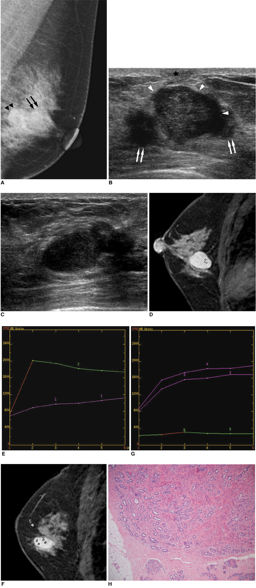
Fig. 1 43-year-old female patient. A. Medio-lateral oblique view of left breast shows two oval isodense masses (arrows and arrowheads) with partially obscured margin (arrows) in left subareolar area. B, C. Ultrasound images show 2.3-cm sized irregular isoechoic mass (white arrowheads) with microlobulated margin located between surgical scars (arrows) and overlying skin scar (black star B), and another, 2.0-cm sized oval hypoechoic mass with circumscribed margin located in lower outer quadrant of left breast (C). D. Contrast-enhanced sagittal subtracted MR image reveals rapidly enhancing mass. E. Graph of MRI time-signal intensity curves from MR image shows rapid enhancement on first postcontrast time point and significant drop in signal on subsequent time points. F. Contrast-enhanced sagittal subtracted MR image reveals progressive enhancement of mass. G. Graph of MRI time-signal intensity curves from MR image shows gradual enhancement on first postcontrast time point and continuous increase in signal on subsequent time points. H. Photomicrograph shows smooth muscle fibers without atypia (Hematoxylin & Eosin stain, ×100).
Reference
-
1. Sharkey FE, Allred DC, Valente PT. Damjanovi I, Linder J, editors. Breast. Anderson's pathology. 1996. St. Louis: Mosby-Yearbook;2363–2364.2. Charpin C, Mathoulin MP, Andrac L, Barberis J, Boulat J, Sarradour B, et al. Reappraisal of breast hamartomas. A morphological study of 41 cases. Pathol Res Pract. 1994. 190:362–371.3. Stafyla V, Kotsifopoulos N, Grigoriadis K, Bakoyiannis CN, Peros G, Sakorafas GH. Myoid hamartoma of the breast: a case report and review of the literature. Breast J. 2007. 13:85–87.4. Di Tommaso L, Pasquinelli G, Damiani S. Smooth muscle cell differentiation in mammary stromo-epithelial lesions with evidence of a dual origin: stromal myofibroblasts and myoepithelial cells. Histopathology. 2003. 42:448–456.5. Ruiz Tovar J, Reguero Callejas ME, Aláez Chillarón AB, Ramiro Pérez C, Collado Guirao MV, Rojo Blanco R, et al. Mammary hamartoma. Clin Transl Oncol. 2006. 8:290–293.6. Georgian-Smith D, Kricun B, McKee G, Yeh E, Rafferty EA, D'Alessandro HA, et al. The mammary hamartoma: appreciation of additional imaging characteristics. J Ultrasound Med. 2004. 23:1267–1273.7. Wiener JI, Schilling KJ, Adami C, Obuchowski NA. Assessment of suspected breast cancer by MRI: a prospective clinical trial using a combined kinetic and morphologic analysis. AJR Am J Roentgenol. 2005. 184:878–886.8. Rosser RJ. Epithelioid cells in myoid hamartoma of the breast. Arch Pathol Lab Med. 1997. 121:354–355.9. Linell F, Ostberg G, Söderström J, Andersson I, Hildell J, Ljungqvist U. Breast hamartomas. An important entity in mammary pathology. Virchows Arch A Pathol Anat Histol. 1979. 383:253–264.
- Full Text Links
-
- Actions
-
Cited
- CITED
-
- Close
- Share
-
- Similar articles
-
- Myoid Hamartoma of the Breast: A Case Report
- Myoid Hamartoma of the Breast: A Case Report
- Myoid Hamartoma of the Breast with Synchronous Contralateral Breast Cancer: Report of a Case
- Invasive Ductal Carcinoma Arising within a Mammary Hamartoma: Case Report
- Recurrent Lipofibromatous Hamartoma of the Median Nerve
