Bilateral Spontaneous Dislocation of Intraocular Lenses within the Capsular Bag in a Retinitis Pigmentosa Patient
- Affiliations
-
- 1Department of Ophthalmology, Kim's Eye Hospital, Myung-Gok Eye Research Institute, Konyang University College of Medicine, Seoul, Korea.
- KMID: 1115784
- DOI: http://doi.org/10.3341/kjo.2004.18.1.52
Abstract
- A 45-year-old man with retinitis pigmentosa (RP), who had undergone uneventful extracapsular cataract extraction (ECCE) in his right eye eight years previously, and phacoemulsification in his left eye six years previously, had spontaneously dislocated intraocular lenses (IOL) within the capsular bag in both eyes one month apart. We removed the dislocated IOLs, and performed anterior vitrectomy and scleral fixation of the new IOLs. Mild contraction of the capsular bags and uneven distribution of the zonular remnants' clumps along the equator of the capsules were found by scanning electron microscopic (SEM) examination. In this study, we propose the correlation between RP and zonular weakness. To our knowledge, this is the first case report of bilateral spontaneous dislocation of IOLs within the capsular bag of an RP patient.
Keyword
MeSH Terms
Figure
-
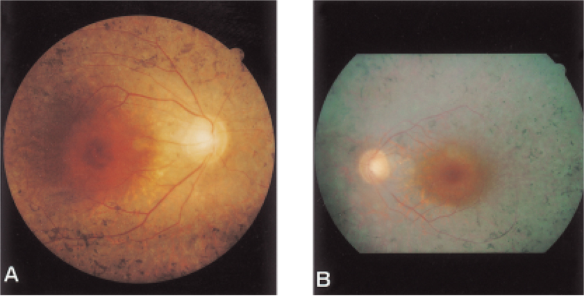
Fig. 1 Fundus photograph of the right(A) and left(B) eyes. Bone-spicule pigmentation just anterior to the posterior pole with normal-appearing macula and slightly pale optic disc are evident.
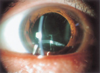
Fig. 2 Slit lamp photograph. IOL dislocation in the right eye after pupil dilation six years after phacoemulsification can be seen.
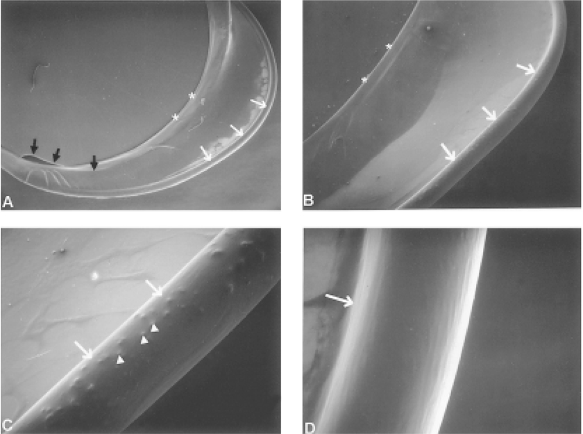
Fig. 3 Scanning electron microscopy. A: The removed intraocular lens within the capsular bag in the right eye with phacoemulsification. B, C: The zonular remnants' clumps are unevenly distributed on the equator. D: There are some parts with no zonules. Equator and haptic, white arrows; continuous curvilinear capsulorhexis margin, asterisks; optic, black arrows; zonular remnants' clumps, arrowheads (A: original magnification ×22. B: original magnification ×44. C: original magnification ×178. D: original magnification ×356).
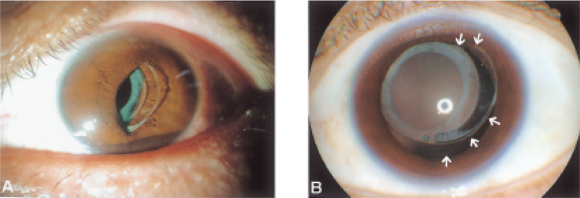
Fig. 4 Slit lamp photograph. A: A haptic has been prolapsed into the anterior chamber and the optic has been captured by the temporal iris in the left eye, but they are still in the bag. B: After pupil dilation, zonular dehiscence can be seen between the positions of 1 to 6 o'clock (white arrows).
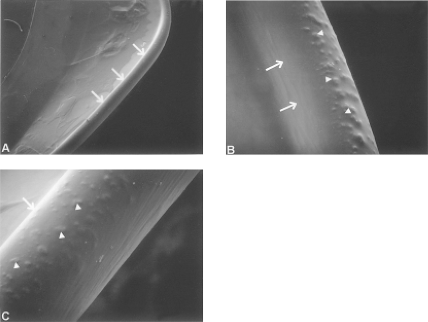
Fig. 5 Scanning electron microscopy. The zonular remnants (arrowheads) are found more evenly distributed and densely on most of the equator (white arrows) of the capsular bag in the left eye which underwent ECCE than the right eye. (A: original magnification ×44. B: original magnification ×178. C: original magnification ×356).
Cited by 1 articles
-
Comparison of Clinical Outcomes after Various Techniques of Intraocular Lens Dislocation Correction
Jae Hong Sun, Jae Yong Kim, Myoung Joon Kim, Young Hee Yoon, Hung Won Tchah
J Korean Ophthalmol Soc. 2014;55(2):196-201. doi: 10.3341/jkos.2014.55.2.196.
Reference
-
1. Jackson H, Garway-Heath D, Rosen P, Bird AC, Tuft SJ. Outcome of cataract surgery in patients with retinitis pigmentosa. Br J Ophthalmol. 2001. 85:936–938.2. Reccia R, Scala A, Bosone G. Posterior chamber intraocular lens implantation in patients with retinitis pigmentosa. Doc Ophthalmol. 1989. 72:115–118.3. Newsome DA, Stark WJ Jr, Maumenee IH. Cataract extraction and intraocular lens implantation in patient with retinitis pigmentosa or Usher's syndrome. Arch Ophthalmol. 1986. 104:852–854.4. Nishi O, Nishi K, Fusisawa T. Effects of cytokines on the proliferation of and collagen synthesis by human cataract lens epithelial cells. Br J Ophthalmol. 1996. 80:63–68.5. Rachipalli R, Srinivas K. Capsulorhexis phimosis in retinitis pigmentosa despite capsular tension ring implantation. J Cataract Refract Surg. 2001. 27:1691–1964.6. Hayashi K, Hayashi H, Matsuo K, Nakao F, Hayashi F. Anterior capsule contraction and intraocular lens dislocation after implant surgery in eyes with retinitis pigmentosa. Ophthalmology. 1998. 105:1239–1243.7. Namiki M, Tagami Y, Morino I. Findings from slit lamp and histological examination of the anterior capsule in patients with severe anterior capsule shrinkage and opacities after implantation of intraocular lens. J Jpn Ophthalmol Soc. 1993. 97:716–720.8. Lee DH, Yoon BJ, Kim HJ. Intraocular lens implantation in retinitis pigmentosa patients. J Korean Ophthalmol Soc. 1990. 31:1520–1522.9. Nishi O, Nishi K. Intraocular lens encapsulation by shrinkage of the capsulorhexis opening. J Cataract Refract Surg. 1993. 19:544–545.10. Shigeeda T, Nagahara M, Kato S, Kunimatsu S, Kaji Y, Tanaka S, Amano S, Oshika T. Spontaneous posterior dislocation of intraocular lenses fixated in the capsular bag. J Cataract Refract Surg. 2002. 28:1689–1693.11. Framme C, Hoerauf H, Laqua H. Delayed intraocular lens dislocation after neodymium: YAG capsulotomy. J Cataract Refract Surg. 1998. 24:1541–1543.
- Full Text Links
-
- Actions
-
Cited
- CITED
-
- Close
- Share
-
- Similar articles
-
- Bilateral Spontaneous Anterior Lens Dislocation in a Retinitis Pigmentosa Patient
- A Case of Unilateral Retinitis Pigmentosa
- A Case of Retinitis Pigmentosa without Pigment
- Clinical Results of Cataract Operations using Piggy-back Method
- The Effect of Capsular Tension Ring on Shape of Capsular Bag, Capsular Opening, and Intraocular Lens
