Management of Dermoid Tumor in the Medial Canthal Area
- Affiliations
-
- 1Department of Ophthalmology, Seoul National University College of Medicine, Seoul, Korea. khwarg@snu.ac.kr
- 2Department of Ophthalmology, Seoul National University Bundang Hospital, Seongnam, Korea.
- 3Department of Ophthalmology, Seoul National University Boramae Hospital, Seoul, Korea.
- KMID: 1115754
- DOI: http://doi.org/10.3341/kjo.2009.23.3.204
Abstract
- Dermoid tumors in the medial canthal area are rare, but when present they commonly adhere to the lacrimal canaliculi. Three patients presented with a mass in the medial canthal area. The authors performed excisional biopsies, and the masses were diagnosed as dermoid tumors. In two patients, canalicular lacerations were found after mass excision, which suggested that the masses had been firmly adherent to the lacrimal canaliculi. The lacerated canaliculi were repaired after bicanalicular silicone intubation. In the remaining patient, lacrimal silicone intubation was performed at the beginning of surgery, and the mass was successfully dissected from the canaliculi, leaving them intact. Excision of dermoid tumors in the medial canthal area requires careful dissection to avoid canalicular laceration. Bicanalicular silicone intubation at the beginning of surgery is helpful for the identification of the canaliculi and for the prevention of canalicular laceration during dermoid tumor excision.
Keyword
MeSH Terms
Figure
-

Fig. 1 Face photograph, orbital CT finding and histopathological features of Patient 1. (A) Note the 1 cm round mass just inferior to the left medial canthal tendon. (B) (A) small non-enhancing or minimally enhancing round lesion (arrow) was found in front of the lacrimal sac, in the left medial canthal area. (C) Hematoxylin and eosin stained histopathological section showed that the cyst was surrounded by stratified squamous epithelium (H&E, ×200).
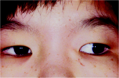
Fig. 2 Face photograph of Patient 2. Note the 1.5 cm round mass in the left medial canthal area.
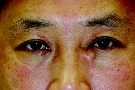
Fig. 3 Face photograph of Patient 3. Note the 1.5 cm round mass just inferior to left medial canthal tendon.
Cited by 1 articles
-
Fibroma of the Medial Canthal Tendon: a Case Report
Jun Woo Chun, Sang Won Hwang
J Korean Ophthalmol Soc. 2013;54(1):155-159. doi: 10.3341/jkos.2013.54.1.155.
Reference
-
1. Rootman J. Disease of the orbit. 2003. 2nd ed. Philadelphia: Lippincott Williams & Wilkins;418–423.2. Hurwitz JJ, Rodgers J, Doucet TW. Dermoid tumor involving the lacrimal drainage pathway: a case report. Ophthalmic Surg. 1982. 13:377–379.3. De La Luz Orozco-Covarrubias MA, Salazar-Leon JA, Tamayo-Sanchez L, et al. Dermoid cyst connected with the lacrimal canaliculum. Pediatr Dermatol. 1993. 10:69–70.
