A Cadaveric Anatomical Study of the Levator Aponeurosis and Whitnall's Ligament
- Affiliations
-
- 1Department of Ophthalmology, College of Medicine, Hanyang University, Hanyang Guri Hospital, Seoul, Korea. lyjot@hanyang.ac.kr
- 2Department of anatomy and cell biology, College of Medicine, Hanyang University, Seoul, Korea.
- KMID: 1115750
- DOI: http://doi.org/10.3341/kjo.2009.23.3.183
Abstract
- PURPOSE
To identify the anatomy of the levator aponeurosis (LA) and Whitnall's ligament (WL) in Korean subjects using cadavers. METHODS: Orbital exenteration was performed in ten cadavers (20 eyeballs) that had no history of trauma near the eyeball. We observed characteristics of WL (tension, density, and shape) and the relationship between the superior rectus muscle (SR) and the levator palpebrae superioris. We measured the distance from both the eyelid margin and the upper border of the tarsal plate to the insertion of the LA medially, centrally, and laterally. RESULTS: The WLs we observed showed several shapes. In 12 eyes, we saw clear, white fibrotic bands, while in four others, we found thin, less taut bands. In four eyes, we were unable to identify the precise shape of the band. The insertions of the LA showed nasal dehiscence in 13 eyes and parallel attachment in seven eyes. The distances from the eyelid margin to the insertion of the LA medially, centrally, and laterally were 8.31 mm, 5.57 mm, and 5.15 mm, respectively. The distances from the upper border of the tarsal plate to the insertion of the LA medially, centrally, and laterally were 2.75 mm, 4.82 mm, and 4.29 mm, respectively. CONCLUSIONS: This study examined the anatomy of WL and the LA in Korean subjects and may be helpful as a reference in levator muscle surgery.
MeSH Terms
Figure
-
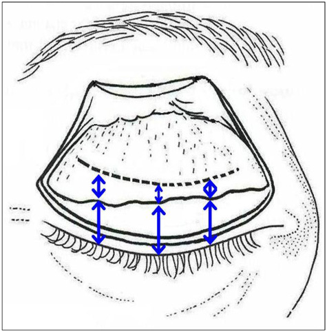
Fig. 1 Drawing showing the measurement of the distance of insertion of the LA from the tarsal plate (short arrows) and from the eyelid margin (long arrows) after dividing the upper eyelid into medial, central, and lateral parts.
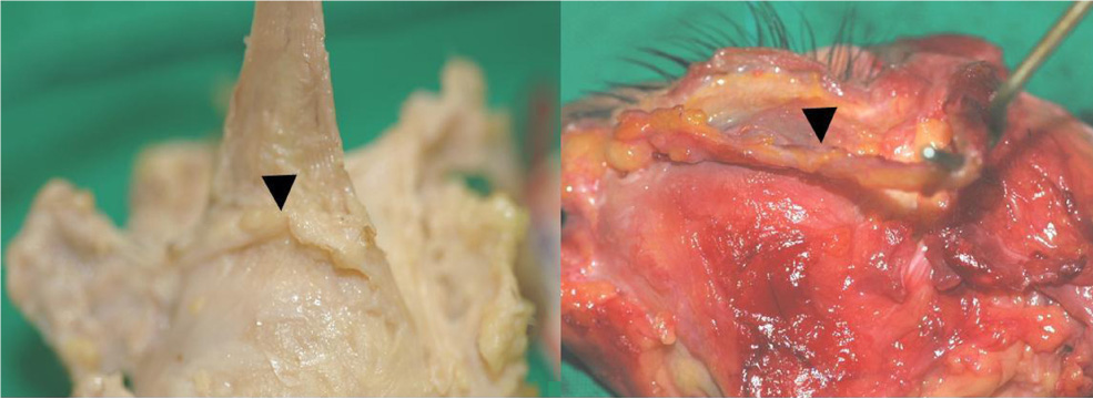
Fig. 2 Photographs showing taut (left) and wide band-shaped (right) Whitnall's ligaments (arrowhead).

Fig. 3 Photographs showing weak, string-shaped Whitnall's ligaments (arrowhead).

Fig. 4 Photographs showing undifferentiated Whitnall's ligaments.
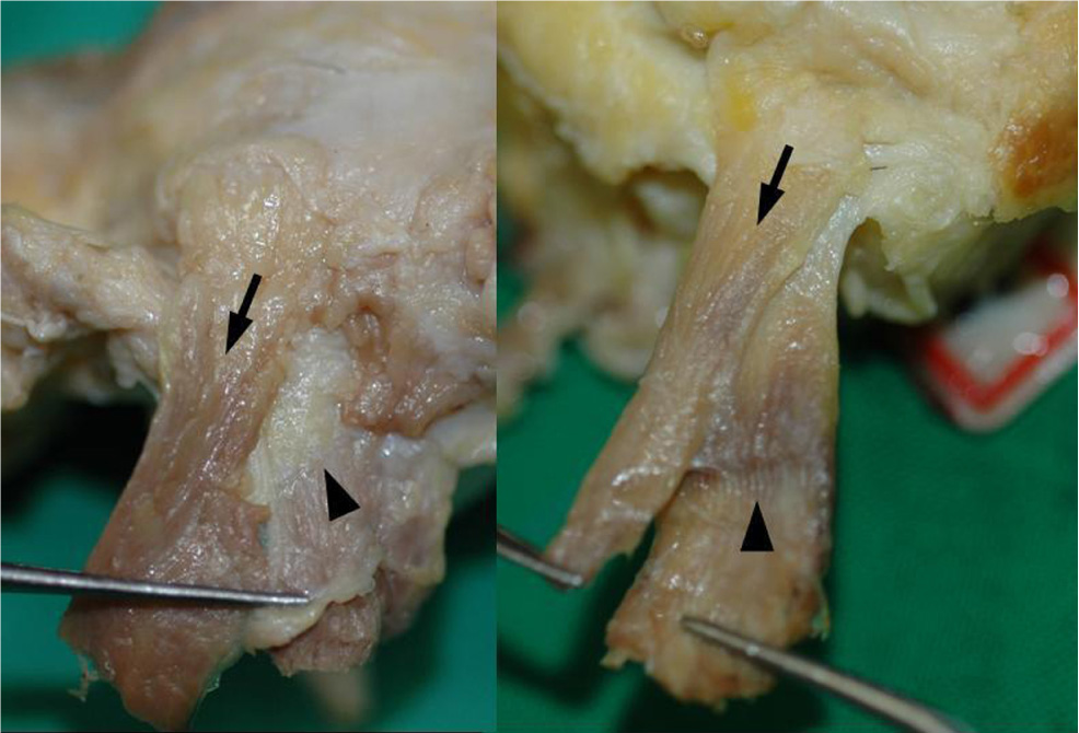
Fig. 5 Photographs showing the X-shaped relationship between the levator palpebrae superioris (arrow) and the superior rectus muscle (arrowhead) from a superior view.
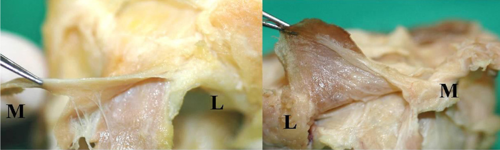
Fig. 6 Photographs showing the medial attachment of the levator palpebrae superioris (upper side muscle)and the superior rectus muscle (lower side muscle) connected by a fibrous membrane (M: medial, L: lateral).
Reference
-
1. Anderson RL, Dixon RS. Aponeurotic ptosis surgery. Arch Ophthalmol. 1979. 97:1123–1128.2. Anderson RL, Beard C. The levator aponeurosis: attachments and their clinical significance. Arch Ophthalmol. 1977. 95:1437–1441.3. Anderson RL, Dixon RS. The role of Whitnall's ligament in ptosis surgery. Arch Ophthalmol. 1979. 97:705–707.4. Whitnall SE. A ligament acting as a check to the action of the levator palpebrae superioris muscle. J Anat Physiol. 1910. 45:131–139.5. Whitnall SE. The Anatomy of the Human Orbit and Accessory Organs of Vision. 1932. 2nd ed. Oxford, England: Humphrey Milford;143–151.6. Fink WH. Anatomical study of the orbital fascia. Trans Am Acad Ophthalmol Otolaryngol. 1959. 63:54–63.7. Lemke BN, Stasior OG, Rosenberg PN. The surgical relations of the levator palpebare superioris muscle. Ophthal Plast Reconstr Surg. 1988. 4:25–30.8. Anderson RL, Jordan DR, Dutton JJ. Whitnall's sling for poor function ptosis. Arch Ophthalmol. 1990. 108:1628–1632.9. Codere F, Tucker NA, Renaldi B. The anatomy of Whitnall ligament. Ophthalmology. 1995. 102:2016–2019.10. Cho YJ, Kim YS, Chung WS. Anatomical structure and its clinical significance of Whitnall's ligament in patients with ptosis. J Korean Ophthalmol Soc. 1996. 37:427–433.11. Jeong S, Lemke BN, Dortzbach RK, et al. The asian upper eyelid: An anatomical study with comparison to the Caucasian eyelid. Arch Ophthalmol. 1999. 117:907–912.12. Haramoto U, Kubo T, Tamatani M, Hosokawa MK. Anatomic study of the insertion of the levator aponeurosis and Muller's muscle in oriental eyelids. Ann Plasy Surg. 2001. 47:528–533.
- Full Text Links
-
- Actions
-
Cited
- CITED
-
- Close
- Share
-
- Similar articles
-
- Anatomical structure and Its Clinical Significance of Whitnall's Ligament in Patients with Ptosis
- Ptosis Correction using Partial Incision Technique
- Use of the Levator Muscle as a Frontalis Sling in Monocular Elevation Deficiency
- Cyst Between the Levator Aponeurosis and the Palpebral Conjunctiva
- Histological Changes in Levator Aponeurosis According to Blepharoptosis and Aging
