Korean J Ophthalmol.
2009 Sep;23(3):176-182. 10.3341/kjo.2009.23.3.176.
Structural and Functional Relationships in Glaucoma Using Standard Automated Perimetry and the Humphrey Matrix
- Affiliations
-
- 1Department of Ophthalmology, University of Pochon, College of Medicine, CHA Medical Center, Bundang, Korea.
- 2Department of Ophthalmology, University of Ulsan, College of Medicine, Asan Medical Center, Seoul, Korea. mskook@amc.seoul.kr
- KMID: 1115749
- DOI: http://doi.org/10.3341/kjo.2009.23.3.176
Abstract
- PURPOSE
To evaluate and compare correlations between structural and functional loss in glaucoma as assessed by optical coherence tomography (OCT), scanning laser polarimetry (GDx VCC, as this was the model used in this study), standard automated perimetry (SAP), and the Humphrey Matrix (Matrix). METHODS: Ninety glaucomatous eyes identified with SAP and 112 eyes diagnosed using Matrix were independently classified into six subgroups, either S1/M1 (MD>-6dB), S2/M2 (-12
Keyword
MeSH Terms
Figure
-
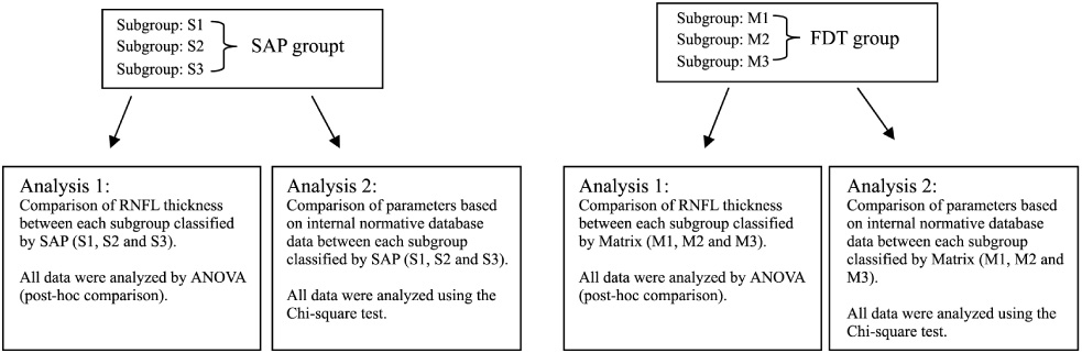
Fig. 1 Overview of the division of the SAP and Matrix groups and Analyses 1 and 2.
Reference
-
1. Kerrgan-Baumrind LA, Quigley HA, Pease ME, et al. Number of ganglion cells in glaucoma eyes compared with threshold visual field tests in the same persons. Invest Ophthalmol Vis Sci. 2000. 41:741–748.2. Quigley HA, Addicks EM, Green WR. Optic nerve damage in human glaucoma. III. Quantitative correlation of nerve fiber loss and visual field defect in glaucoma, ischemic neuropathy, papilledema, and toxic neuropathy. Arch Ophthalmol. 1982. 100:135–146.3. Quigley HA, Dunkelberger GR, Green WR. Retinal ganglion cell atrophy correlated with automated perimetry in human eyes with glaucoma. Am J Ophthalmol. 1989. 107:453–464.4. Harwerth RS, Quigley HA. Visual field defects and retinal ganglion cell losses in patients with glaucoma. Arch Ophthalmol. 2006. 124:853–859.5. Maddess T, Goldberg I, Dobinson J, et al. Testing for glaucoma with the spatial frequency doubling illusion. Vision Res. 1999. 39:4258–4273.6. Soliman MA, de Jong LA, Ismaeil AA, et al. Standard achromatic perimetry, short wavelength automated perimetry, and frequency doubling technology for detection of glaucoma damage. Ophthalmology. 2002. 109:444–454.7. Kogure S, Toda Y, Tsukahara S. Prediction of future scotoma on conventional automated static perimetry using frequency doubling technology perimetry. Br J Ophthalmol. 2006. 90:347–352.8. Medeiros FA, Sample PA, Weinreb RN. Frequency doubling technology perimetry abnormalities as predictors of glaucomatous visual field loss. Am J Ophthalmol. 2004. 137:863–871.9. Kamantigue MEG, Joson PJ, Chen PP. Prediction of visual field defects on standard automated perimetry by screening C-20-1 frequency doubling technology perimetry. J Glaucoma. 2006. 15:35–39.10. Huang XR, Knighton RW. Linear birefringence of the retinal nerve fiber layer measured in vitro with a multispectral imaging micropolarimeter. J Biomed Opt. 2002. 7:199–204.11. Dreher AW, Bailey ED. Assessment of the retinal nerve fiber layer by scanning-laser polarimetry. SPIE. 1993. 1877:266–271.12. Zangwill LM, Medeiros FA, Bowd C, Weinreb RN. Grehn F, Stamper R, editors. Optic nerve imaging: recent advances. Glaucoma. 2004. Vol.2. Berlin: Springer Verlag;63–91.13. Reus NJ, Colen TP, Jemij HG. Visualization of localized retinal nerve fiber layer defects with the GDx with individualized and with fixed compensation of anterior segment birefringence. Ophthalmology. 2003. 110:1512–1516.14. Huang D, Swanson EA, Lin CP, et al. Optical coherence tomography. Science. 1991. 254:1178–1181.15. Izatt JA, Hee MR, Swanson EA, et al. Micrometer-scale resolution imaging of the anterior eye in vivo with optical coherence tomography. Arch Ophthalmol. 1994. 112:1584–1589.16. Schuman JS, Hee MR, Puliafito CA, et al. Quantification of nerve fiber layer thickness in normal and glaucomatous eyes using optical coherence tomography. Arch Ophthalmol. 1995. 113:586–596.17. Kass MA, Heuer DK, Higginbotham EJ, et al. The Ocular Hypertension Treatment Study: a randomized trial determines that topical ocular hypotensive medication delays or prevents the onset of primary open-angle glaucoma. Arch Ophthalmol. 2002. 120:701–713.18. European Glaucoma Prevention Study (EGPS) Group. Results of the European Glaucoma Prevention Study. Ophthalmology. 2005. 112:366–375.19. Zhou Q, Weinreb RN. Individualized compensation of anterior segment birefringence during scanning laser polarimetry. Invest Ophthalmol Vis Sci. 2002. 43:2221–2228.20. Weinreb RN, Bowd C, Zangwill LM. Glaucoma detection using scanning laser polarimetry with variable corneal polarization compensation. Arch Ophthalmol. 2003. 121:218–224.21. Hodapp E, Parrish RK, Anderson DR. Clinical decisions in glaucoma. 1993. Vol.1. St. Louis: Mosby;52–63.22. Beck RW, Bergstrom TJ, Lichter PR. A clinical comparison of visual field testing with a new automated perimeter, the Humphrey Field Analyzer and the Goldmann perimeter. Ophthalmology. 1985. 92:77–82.23. Sample PA, Bosworth CF, Blumenthal EZ, et al. Visual functionspecificperimetry for indirect comparison of different ganglioncell populations in glaucoma. Invest Ophthalmol Vis Sci. 2000. 41:1783–1790.24. Harwerth R, Carter-Dawson L, Shen F, et al. Ganglion cell losses underlying visual field defects from experimental glaucoma. Invest Ophthalmol Vis Sci. 1999. 40:2242–2250.25. White AJ, Sun H, Swanson WH, Lee BB. An examination of physiological mechanisms underlying the frequency-doubling illusion. Invest Ophthalmol Vis Sci. 2002. 43:3590–3599.26. Cello KE, Nelson-Quigg JM, Johnson CA. Frequency doubling technology perimetry for detection of glaucomatous visual field loss. Am J Ophthalmol. 2000. 129:314–322.27. Johnson CA, Samuels SJ. Screening for glaucomatous visual field loss with frequency-doubling perimetry. Invest Ophthalmol Vis Sci. 1997. 38:413–425.28. Quigley HA. Identification of glaucoma-related visual field abnormality with the screening protocol of frequency doubling technology. Am J Ophthalmol. 1998. 125:819–829.29. Maddess T, Henry GH. Performance of nonlinear visual units in ocular hypertension and glaucoma. Clin Vis Sci. 1992. 7:371–383.30. Brusini P, Busatto P. Frequency doubling perimetry in glaucoma early diagnosis. Acta Ophthalmol Scand. 1998. 76(S227):23–24.31. Burnstein Y, Ellish NJ, Magbalon M, et al. Comparison of frequency doubling perimetry with Humphrey visual field analysis in a glaucoma practice. Am J Ophthalmol. 2000. 129:328–333.32. Johnson CA, Samuels JS. Screening for glaucomatous visual field loss with frequency-doubling perimetry. Invest Ophthalmol Vis Sci. 1997. 38:413–425.33. Patel SC, Friedman DS, Varadkar P, et al. Algorithm for interpreting the results of frequency doubling perimetry. Am J Ophthalmol. 2000. 129:323–327.34. Sample PA, Medeiros FA, Racette L, et al. Identifying glaucomatous vision loss with visual-function-specific perimetry in the diagnostic innovations in glaucoma study. Invest Ophthalmol Vis Sci. 2006. 47:3381–3389.35. Paczka JA, Friedman DS, Quigley HA, et al. Diagnostic capabilities of frequency doubling technology, scanning laser polarimetry and nerve fiber layer photographs to distinguish glaucomatous damage. Am J Ophthalmol. 2001. 131:188–197.36. Serguhn S, Spiegel D. Comparison of frequency doubling perimetry and standard achromatic computerized perimetry in patients with glaucoma. Graefes Arch Clin Exp Ophthalmol. 2001. 239:351–355.37. Fukushima A, Shirakashi M, Yaoeda K, et al. Relationship between indices of Humphrey perimetry and frequency doubling technology perimetry in glaucoma. J Glaucoma. 2004. 13:114–119.38. Sponsel WE, Arango S, Trigo Y, et al. Clinical classification of glaucomatous visual field loss by frequency doubling perimetry. Am J Ophthalmol. 1998. 125:830–836.39. Medeiros FA, Zangwill LM, Bowed C, et al. Fourier analysis of scanning laser polarimetry measurements with variable corneal compensation in glaucoma. Invest Ophthalmol Vis Sci. 2003. 44:2606–2612.40. Medeiros FA, Zangwill LM, Bowed C, et al. Evaluation of retinal nerve fiber layer, optic nerve head, and macular thickness measurements for glaucoma detection using optical coherence tomopraphy. Am J Ophthalmol. 2005. 139:44–55.41. Budenz DL, Michael A, Chang RT, et al. Sensitivity and specificity of the Stratus OCT for perimetric glaucoma. Ophthalmology. 2005. 112:3–9.42. Bagga H, Greenfield DS, Feuer W, et al. Scanning Laser Polarimetry With Variable Corneal Compensation and Optical Coherence Tomography in Normal and Glaucomatous Eyes. Am J Ophthalmol. 2003. 135:521–529.43. Kanamori A, Nakamura M, Escano MFT, et al. Evaluation of the Glaucomatous Damage on Retinal nerve fiber layer thickness measured by Optical Coherence Tomography. Am J Ophthalmol. 2003. 135:513–520.44. Blumenthal EZ, Williams JM, Weinreb RN, et al. Reproducibility of nerve fiber layer thickness measurements by use of optical coherence tomography. Ophthalmology. 2000. 107:2278–2282.45. Carpineto P, Ciancaglini M, Zuppardi E, et al. Reliability of nerve fiber layer thickness measurements using optical coherence tomography in normal and glaucomatous eyes. Ophthalmology. 2003. 110:190–195.46. Kook MS, Sung K, Park RH, et al. Reproducibility of scanning laser polarimetry (GDx) of peripapillary retinal nerve fiber layer thickness in normal subjects. Graefes Arch Clin Exp Ophthalmol. 2001. 239:118–121.47. Reus NJ, Nemij HG. The relationship between standard automated perimetry and GDx VCC measurements. Invest Ophthalmol Vis Sci. 2004. 45:840–845.48. Kim SH, Yang SJ, Kook MS, et al. Correlation Between Frequency Doubling Technology Perimetry and Scanning Laser Polarimetry in Glaucoma Suspects and Glaucomatous Eyes. Korean J ophthalmol. 2004. 18:89–99.
- Full Text Links
-
- Actions
-
Cited
- CITED
-
- Close
- Share
-
- Similar articles
-
- The Comparison of The Matrix Perimetry and Humphrey Standard Perimetry in Various Patients Group
- Performance of Humphrey Matrix Frequency Doubling Technology Perimetry and Standard Automated Perimetry Global Indices
- Usefulness of the Binocular Double Vision Field Using Kinetic Automated Perimetry in Diplopia
- Comparison of the Normal Visual Fields Between the Goldmann and Humphrey Kinetic Perimetries
- Comparison of Total Deviation of Standard Automated Perimetry and Matrix FDT in Preperimetric Glaucoma Patients
