Scanning Laser Polarimetry and Optical Coherence Tomography for Detection of Retinal Nerve Fiber Layer Defects
- Affiliations
-
- 1Department of Ophthalmology, National Medical Center, Seoul, Korea.
- 2Department of Ophthalmology, Korea University College of Medicine, Seoul, Korea. yongykim@mail.korea.ac.kr
- KMID: 1115748
- DOI: http://doi.org/10.3341/kjo.2009.23.3.169
Abstract
- PURPOSE
To compare the ability of scanning laser polarimetry with variable corneal compensation (GDx-VCC) and Stratus optical coherence tomography (OCT) to detect photographic retinal nerve fiber layer (RNFL) defects. METHODS: This retrospective cross-sectional study included 45 eyes of 45 consecutive glaucoma patients with RNFL defects in red-free fundus photographs. The superior and inferior temporal quadrants in each eye were included for data analysis separately. The location and presence of RNFL defects seen in red-free fundus photographs were compared with those seen in GDx-VCC deviation maps and OCT RNFL analysis maps for each quadrant. RESULTS: Of the 90 quadrants (45 eyes), 31 (34%) had no apparent RNFL defects, 29 (32%) had focal RNFL defects, and 30 (33%) had diffuse RNFL defects in red-free fundus photographs. The highest agreement between GDx-VCC and red-free photography was 73% when we defined GDx-VCC RNFL defects as a cluster of three or more color-coded squares (p<5%) along the traveling line of the retinal nerve fiber in the GDx-VCC deviation map (kappa value, 0.388; 95% confidence interval (CI), 0.195 to 0.582). The highest agreement between OCT and red-free photography was 85% (kappa value, 0.666; 95% CI, 0.506 to 0.825) when a value of 5% outside the normal limit for the OCT analysis map was used as a cut-off value for OCT RNFL defects. CONCLUSIONS: According to the kappa values, the agreement between GDx-VCC deviation maps and red-free photography was poor, whereas the agreement between OCT analysis maps and red-free photography was good.
Keyword
MeSH Terms
Figure
-

Fig. 1 Topographic measurements of focal retinal nerve fiber layer (RNFL) defects in the same eye. In this eye, the RNFL defect in the inferior temporal quadrant was detected by both GDx-VCC and OCT. However, the RNFL defect in the superior temporal quadrant was detected by only OCT. (A) Topographic measurements of RNFL defects in a red-free photograph. The reference line (line r) is the horizontal line temporal to the disc center (c). Lines a (a') and b (b') are the lines from the center of the disc tothe disc margin where the RNFL defect meets the disc. The minimum and maximum angles between line r and the lines a (a') or b (b'), respectively, were measured. The arrows indicate focal RNFL defects seen in the red-free photograph. (B) Overlap between the GDx-VCC deviation map and the red-free photographs in the inferior quadrant. (C) Topographic measurements of RNFL defects in the OCT RNFL analysis map. The minimum and maximum angles of the line graph segments located below the yellow band were measured after 256 tests points were converted to 360 degrees of the angle.

Fig. 2 Photographs showing diffuse retinal nerve fiber layer (RNFL) defects detected in the same eye. The red-free photograph shows diffuse RNFL defects in the superior temporal quadrant (A). A decreased intensity in the upper half compared with the intensity in the lower half was noted. The location of RNFL defects in the GDx-VCC deviation map (B) and the OCT RNFL analysis map (C) overlap with diffuse RNFL defects seen in the red-free photograph.

Fig. 3 Photographs showing various definitions of the deviation map using scanning laser polarimetry (GDx-VCC). A to D. No GDx retinal nerve fiber layer (RNFL) defect is shown in the deviation maps (A: one square, B: two squares, C: squares outside one disc diameter, and D: nasal squares). E to L. Several GDx RNFL defects are shown in the deviation maps (please refer to the methods section in the text).
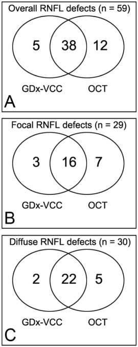
Fig. 4 Venn diagrams showing agreements for overall (A), focal (B), and diffuse (C) photographic retinal nerve layer defects (RNFL) with scanning laser polarimetry (GDx-VCC) and optical coherence tomography (OCT).
Cited by 2 articles
-
Peripapillary Retinal Nerve Fiber Layer Thicknesses Did Not Change in Long-term Hydroxychloroquine Users
Eun Jung Lee, Sang Jin Kim, Jong Chul Han, Doo Ri Eo, Min Gyu Lee, Don-Il Ham, Se Woong Kang, Changwon Kee, Jaejoon Lee, Hoon-Suk Cha, Eun-Mi Koh
Korean J Ophthalmol. 2018;32(6):459-469. doi: 10.3341/kjo.2018.0004.Influence of Epiretinal Membranes on the Retinal Nerve Fiber Layer Thickness Measured by Spectral Domain Optical Coherence Tomography in Glaucoma
Ju Mi Kim, Kyoung Nam Kim, Woo-Jin Kim, Chang-sik Kim
Korean J Ophthalmol. 2019;33(5):422-429. doi: 10.3341/kjo.2018.0105.
Reference
-
1. Harwerth RS, Carter-Dawson L, Shen F, et al. Ganglion cell losses underlying visual field defects from experimental glaucoma. Invest Ophthalmol Vis Sci. 1999. 40:2242–2250.2. Sommer A, Katz J, Quigley HA, et al. Clinically detectable nerve fiber atrophy precedes the onset of glaucomatous field loss. Arch Ophthalmol. 1991. 109:77–83.3. Airaksinen PJ, Drance SM, Douglas GR, et al. Visual field and retinal nerve fiber layer comparisons in glaucoma. Arch Ophthalmol. 1985. 103:205–207.4. Quigley HA, Katz J, Derick RJ, et al. An evaluation of optic disc and nerve fiber layer examinations in monitoring progression of early glaucoma damage. Ophthalmology. 1992. 99:19–28.5. Quigley HA, Miller NR, George T. Clinical evaluation of nerve fiber layer atrophy as an indicator of glaucomatous optic nerve damage. Arch Ophthalmol. 1980. 98:1564–1571.6. Sommer A, Quigley HA, Robin AL, et al. Evaluation of nerve fiber layer assessment. Arch Ophthalmol. 1984. 102:1766–1771.7. Tuulonen A, Airaksinen PJ. Initial glaucomatous optic disk and retinal nerve fiber layer abnormalities and their progression. Am J Ophthalmol. 1991. 111:485–490.8. Niessen AG, van den Berg TJ, Langerhorst CT, Bossuyt PM. Grading of retinal nerve fiber layer with a photographic reference set. Am J Ophthalmol. 1995. 120:577–586.9. Weinreb RN, Shakiba S, Zangwill L. Scanning laser polarimetry to measure the nerve fiber layer of normal and glaucomatous eyes. Am J Ophthalmol. 1995. 119:627–636.10. Greenfield DS, Knighton RW, Feuer WJ, et al. Correction for corneal polarization axis improves the discriminating power of scanning laser polarimetry. Am J Ophthalmol. 2002. 134:27–33.11. Zhou Q, Weinreb RN. Individualized compensation of anterior segment birefringence during scanning laser polarimetry. Invest Ophthalmol Vis Sci. 2002. 43:2221–2228.12. Huang D, Swanson EA, Lin CP, et al. Optical coherence tomography. Science. 1991. 54:1178–1181.13. Schuman JS, Hee MR, Puliafito CA, et al. Quantification of nerve fiberlayer thickness in normal and glaucomatous eyes using optical coherence tomography. Arch Ophthalmol. 1995. 113:586–596.14. Medeiros FA, Zangwill LM, Bowd C, et al. Evaluation of retinal nerve fiberlayer, optic nerve head and macular thickness measurements for glaucoma detection using optical coherence tomography. Am J Ophthalmol. 2005. 139:44–55.15. Bagga H, Greenfield DS, Feuer W, Knighton RW. Scanning laser polarimetry with variable corneal compensation and optical coherence tomography in normal and glaucomatous eyes. Am J Ophthalmol. 2003. 135:521–529.16. Brusini P, Salvetat ML, Zeppieri M, et al. Comparison between GDx VCC scanning laser polarimetry and Stratus OCT optical coherence tomography in the diagnosis of chronic glaucoma. Acta Ophthalmol Scand. 2006. 84:650–655.17. Medeiros FA, Zangwill LM, Bowd C, Weinreb RN. Comparison of the GDx VCC scanning laser polarimeter, HRT II confocal scanning laser ophthalmoscope, and stratus OCT optical coherence tomograph for the detection of glaucoma. Arch Ophthalmol. 2004. 122:827–837.18. Kanamori A, Nagai-Kusuhara A, Escano MF, et al. Comparison of confocal scanning laser ophthalmoscopy, scanning laser polarimetry and optical coherence tomography to discriminate ocular hypertension and glaucoma at an early stage. Graefes Arch Clin Exp Ophthalmol. 2006. 244:58–68.19. Leung CK, Chan W, Chong KKL, et al. Comparative study of retinal nerve fiber layer measurement by StratusOCT and GDx VCC, I: correlation analysis in glaucoma. Invest Ophthalmol Vis Sci. 2005. 46:3214–3220.20. Jeoung JW, Park KH, Kim TW, et al. Diagnostic Ability of Optical Coherence Tomography with a Normative Database to Detect Localized Retinal Nerve Fiber Layer Defects. Ophthalmology. 2005. 112:2157–2163.21. Laser Diagnostic Technologies, Inc.RNFL analysis with GDx VCC, a primer and clinical guide. 2004. Laser Diagnostic Technologies, Inc.;31.22. Kook MS, Cho HS, Seong M, Choi J. Scanning laser polarimetry using variable corneal compensation in the detection of glaucoma with localized visual field defects. Ophthalmology. 2005. 112:1970–1978.23. Choi J, Cho HS, Lee CW, Kook MS. Scanning laser polarimetry with variable corneal compensation in the area of apparently normal hemifield in eyes with normal-tension glaucoma. Ophthalmology. 2006. 113:1954–1960.24. Bagga H, Greenfiel DS, Feuer WJ. Quantitative assessment of atypical birefringence images using scanning laser polarimetry with variable corneal compensation. Am J Ophthalmol. 2005. 139:437–446.25. Airaksinen PJ, Nieminen H. Retinal nerve fiber layer photography in glaucoma. Ophthalmology. 1985. 92:877–879.26. Chihara E, Tanihara H. Parameters associated with papillomacular bundle defect in glaucoma. Graefes Arch Clin Exp Ophthalmol. 1992. 200:511–517.27. Hwang JM, Kim TW, Park KH, et al. Correlation between topographic profiles of localized retinal nerve fiber layer defects as determined by optical coherence tomography and red-free fundus photography. J Glaucoma. 2006. 15:223–228.28. Fleiss JL. Statistical methods for rates and proportions. 1981. 2nd ed. New York: John Wiley and Sons;212–213.29. Jonas JB, Schiro D. Localised wedge shaped defects of the retinal nerve fibre layer in glaucoma. Br J Ophthalmol. 1994. 78:285–290.30. Jonas JB, Nguyen NX, Naumann GOH. The retinal nerve fiber layer in normal eyes. Ophthalmology. 1989. 96:627–632.31. Jonas JB, Dichtl A. Evaluation of the retinal nerve fiber layer. Surv Ophthalmol. 1996. 40:369–378.32. Quigley HA, Addicks EM. Quantitative studies of retinal nerve fiber layer defects. Arch Ophthalmol. 1982. 100:807–814.
- Full Text Links
-
- Actions
-
Cited
- CITED
-
- Close
- Share
-
- Similar articles
-
- Biometry of Retinal Nerve Fiber Layer Thickness by NFA
- The Relationship between the Duration of IOP Elevation during LASIK and Nerve Fiber Layer Thickness Measured by GDx(R)
- Comparison of Stratus OCT and GDx VCC in Detecting Localized Retinal Nerve Fiber Layer Defects
- The Relationship between Optical Coherence Tomography and Scanning Laser Polarimetry Measurements in Glaucoma
- Changes in Macular Retinal Layers and Peripapillary Nerve Fiber Layer Thickness after 577-nm Pattern Scanning Laser in Patients with Diabetic Retinopathy
