A Comparison of 2-Octyl Cyanoacrylate Adhesives versus Conventional Suture Materials for Eyelid Wound Closure in Rabbits
- Affiliations
-
- 1Department of Ophthalmology, Dong-A University College of Medicine, Busan, Korea. hbahn@dau.ac.kr
- 2Department of Pathology, Dong-A University College of Medicine, Busan, Korea.
- KMID: 1111874
- DOI: http://doi.org/10.3341/kjo.2011.25.2.121
Abstract
- PURPOSE
To evaluate the clinical efficacy and histopathological tolerance of 2-octyl cyanoacrylate versus conventional suture materials for eyelid wound closure in rabbits.
METHODS
We performed an experimental study on 16 eyes of eight New Zealand albino rabbits. Eyelid incisions of 15 mm were done 4mm from the upper eyelid margin in both eyes. The eyes of the rabbits were divided into two groups: eyelid incisions of the right eye were closed by a 2-octyl cyanoacrylate adhesive (group A) and eyelid incisions of the left eye were closed by 7-0 nylon sutures (group B). At 1, 2, 4, and 8 weeks after surgery, the rabbits were macroscopically examined and then sacrificed. The specimens of their eyelid tissues were stained by a hematoxylin and eosin stain and Masson-trichrome stain, and were observed under microscope.
RESULTS
Both eyelid surgical closure methods were found to be equally efficacious in fixing the eyelids of groups A and B, and their clinical efficacy was similar. Histopathological findings of the hematoxylin and eosin stain of group A showed less inflammatory infiltration than group B at 2 weeks. There were no significant histopathological differences between the two groups at 1, 4, and 8 weeks. The degree of fibrosis of the Masson-trichrome stain was similar between the two groups at 8 weeks.
CONCLUSIONS
The 2-octyl cyanoacrylate adhesive proved to be an effective eyelid closure method and was very well tolerated by the skin surface. 2-Octyl cyanoacrylate could be used as an alternative tissue adhesive for eyelid wound closure along with conventional suture materials.
MeSH Terms
Figure
-
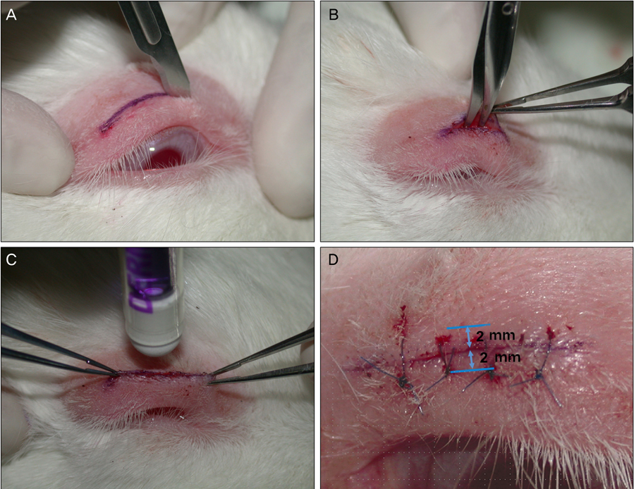
Fig. 1 Surgical technique. (A) A eyelid incision of approximately 15 mm was made. (B) The incision was blotted dry using cotton swabs. Subcutaneous tissue was dissected with the use of Wescott scissors. (C) In group A, a thin layer of 2-octylcyanoacrylate was applied along the edges of the incision. (D) In group B, 7-0 nylon sutures were used in the form of stitches that fixed the edges of the eyelid. Full-thickness biopsies including 2 mm above and below the the incision site were taken for blinded histopathological evaluation. In group A, full-thickness biopsies were also taken by the same method.

Fig. 2 Macroscopic aspect of specimens. Macroscopic aspect of specimens treated with 2-octylcyanoacrylate (A, 1 week; C, 2 weeks) and 7-0 nylon sutures (B, 1 week; D, 2 weeks).

Fig. 3 Histological findings of the incised eyelid (hematoxylin and eosin stain, ×200). (A) The eyelid at 1 week of group A exhibits mild to moderate acute inflammatory cell infiltration and mild to moderate fibroblastic proliferation. (B) The eyelid at 1 week of group B shows moderate acute inflammatory cell infiltration and mild to moderate fibroblastic proliferation and foreign body reaction. (C) The eyelid at 4 weeks of group A shows mild inflammatory cell infiltration. (D) The eyelid at 4 weeks of group B shows mild eosinophilic infiltration. (E) The eyelid at 8 weeks of group A shows nearly normalized morphology with very mild inflammatory cells and fibroblasts. (F) The eyelid at 8 weeks of group B shows mild foreign body inflammatory reaction with the presence of eosinophils.

Fig. 4 Histological findings of fibrosis (Masson-trichrome stain, ×100). (A) The eyelid at 1 week show nearly normalized fibrosis in group A. (B) The eyelid at 1 week show slightly edematous change in group B. The eyelid at 8 weeks show similar findings in group A (C) and group B (D). Mild fibrosis with deposition of collagen is seen.
Reference
-
1. Toriumi DM, O'Grady K. Surgical tissue adhesives in otolaryngology-head and neck surgery. Otolaryngol Clin North Am. 1994. 27:203–209.2. Straatsma BR, Allen RA, Hale PN, Gomez R. Experimental studies employing adhesive compounds in ophthalmic surgery. Trans Am Acad Ophthalmol Otolaryngol. 1963. 67:320–334.3. Ellis RA, Levine AM. Experimental sutureless ocular surgery. Am J Ophthalmol. 1963. 55:733–742.4. Erbil H, Sinav S, Süllü Y, Kandemir B. An experimental study on the use of fibrin sealants in strabismus surgery. Turk J Pediatr. 1991. 33:111–116.5. Leonard F, Kulkarni RK, Brandes G, et al. Synthesis and degradation of poly (alkyl α-cyanoacrylates). J Appl Polym Sci. 1966. 10:259–272.6. Toriumi DM, Raslan WF, Friedman M, Tardy ME. Histotoxicity of cyanoacrylate tissue adhesives. A comparative study. Arch Otolaryngol Head Neck Surg. 1990. 116:546–550.7. Maw JL, Quinn JV, Wells GA, et al. A prospective comparison of octylcyanoacrylate tissue adhesive and suture for the closure of head and neck incisions. J Otolaryngol. 1997. 26:26–30.8. Bernard L, Doyle J, Friedlander SF, et al. A prospective comparison of octyl cyanoacrylate tissue adhesive (dermabond) and suture for the closure of excisional wounds in children and adolescents. Arch Dermatol. 2001. 137:1177–1180.9. Nahas FX, Solia D, Ferreira LM, Novo NF. The use of tissue adhesive for skin closure in body contouring surgery. Aesthetic Plast Surg. 2004. 28:165–169.10. Singer AJ, Hollander JE, Valentine SM, et al. Stony Brook Octylcyanoacrylate Study Group. Prospective, randomized, controlled trial of tissue adhesive (2-octylcyanoacrylate) vs standard wound closure techniques for laceration repair. Acad Emerg Med. 1998. 5:94–99.11. Fogle JA, Kenyon KR, Foster CS. Tissue adhesive arrests stromal melting in the human cornea. Am J Ophthalmol. 1980. 89:795–802.12. Leahey AB, Gottsch JD, Stark WJ. Clinical experience with N-butyl cyanoacrylate (Nexacryl) tissue adhesive. Ophthalmology. 1993. 100:173–180.13. Lee JY, Kim BY, Kim TY. The results of cyanoacrylate glue application for corneal perforation and impending perforation. J Korean Ophthalmol Soc. 2003. 44:2735–2741.14. Bloomfield S, Barnert AH, Kanter PD. The use of Eastman 910 monomer as an adhesive in ocular surgery. I. Biologic effects on ocular tissues. Am J Ophthalmol. 1963. 55:742–748.15. Hong MA, Rapuano CJ. Krachmer JH, Mannis MJ, Holland EJ, editors. Management of corneal perforations. Cornea. 1997. St Louis: Mosby;1818.16. Kim YM, Gupta BK. 2-Octyl cyanoacrylate adhesive for conjunctival wound closure in rabbits. J Pediatr Ophthalmol Strabismus. 2003. 40:152–155.17. Coover HN, Joyner FB, Sheerer NH, et al. Chemistry and performance of cyanoacrylate adhesive. Special Tech Papers. 1959. 5:413–417.18. Pani KC, Gladieux G, Brandes G, et al. The degradation of n-butyl alpha-cyanoacrylate tissue adhesive. II. Surgery. 1968. 63:481–489.19. Refojo MF. Current status of biomaterials in ophthalmology. Surv Ophthalmol. 1982. 26:257–265.20. Alió JL, Gómez J, Mulet E, et al. A new acrylic tissue adhesive for conjunctival surgery: experimental study. Ophthalmic Res. 2003. 35:306–312.21. Quinn J, Maw J, Ramotar K, et al. Octylcyanoacrylate tissue adhesive versus suture wound repair in a contaminated wound model. Surgery. 1997. 122:69–72.22. Quinn JV, Osmond MH, Yurack JA, Moir PJ. N-2-butylcyanoacrylate: risk of bacterial contamination with an appraisal of its antimicrobial effects. J Emerg Med. 1995. 13:581–585.23. Quinn J, Lowe L, Mertz M. The effect of a new tissue-adhesive wound dressing on the healing of traumatic abrasions. Dermatology. 2000. 201:343–346.24. Davis SC, Eaglstein WH, Cazzaniga AL, Mertz PM. An octyl-2-cyanoacrylate formulation speeds healing of partial-thickness wounds. Dermatol Surg. 2001. 27:783–788.25. Singer AJ, Nable M, Cameau P, et al. Evaluation of a new liquid occlusive dressing for excisional wounds. Wound Repair Regen. 2003. 11:181–187.
- Full Text Links
-
- Actions
-
Cited
- CITED
-
- Close
- Share
-
- Similar articles
-
- Comparison of 2-Octylcyanoacrylate and Suture with 8-0 Polyglactin for Conjunctival Wound Closure in Rabbits
- Comparing intra-oral wound healing after alveoloplasty using silk sutures and n-butyl-2-cyanoacrylate
- 2-Octyl Cyanoacrylate Glue Application on Split-Thickness Skin Graft for Lower Extremity Reconstruction
- Analysis of Octyl-2-Cyanoacrylate as a Dressing Material after Pediatric Urological Procedures
- Comparing Conventional Suture Method Versus Wound Closure Using Tissue Glue(Histoacryl Blue(R)): a Prospective Randomized Clinical Trial
