J Vet Sci.
2007 Dec;8(4):427-429. 10.4142/jvs.2007.8.4.427.
A Gartner duct cyst of the vagina causing dysuria and dyschezia in a Yorkshire Terrier
- Affiliations
-
- 1Haemaru Referral Animal Hospital, Sungnam 463-050, Korea. vetboy@netsgo.com
- 2Veterinary Medical Teaching Hospital, College of Veterinary Medicine, Seoul National University, Seoul 151-742, Korea.
- KMID: 1106225
- DOI: http://doi.org/10.4142/jvs.2007.8.4.427
Abstract
- A 5 year-old, intact female Yorkshire terrier was referred for dysuria and dyschezia. The radiographic and ultrasound examination showed a round shaped mass caudal to the urinary bladder that contained anechoic fluid within the thin walls. During surgery, the cyst was noted to be attached to the outer wall of the vagina, not connected to the vaginal lumen. Cystic fluid was removed and the cystic wall was resected. Then the remaining cystic wall was omentalized to prevent a recurrence. Histological examination confirmed that the cyst was of Wolffian duct origin. In this case, a large Gartner duct cyst causing urological problems was diagnosed and removed by surgical resection.
Keyword
MeSH Terms
Figure
-
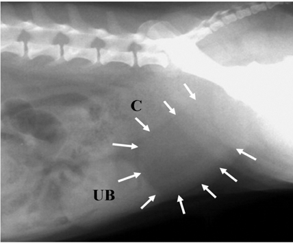
Fig. 1 Right lateral view of abdominal radiography. There was a soft tissue density mass (arrows) caudal to urinary bladder (UB). There was normal colon (C).

Fig. 2 Caudal obdominal ultrasonography. There was a cyst (long arrows) different form urinary bladder (UB) and uterus (short arrows).

Fig. 3 (A) Cyst was located caudal to urinary bladder (UB) and uterus (U). The cyst attached outer wall of cranial vagina. (B) There was no connecting between cystic wall (*) and vaginal lumen.

Fig. 4 (A) Microphotographs of the wall of Gartner duct cyst. Low columnar epithelial cell with smooth muscle in the lamina propria. H&E stain, ×100. (B) High magnification. H&E stain, ×1,000.
Reference
-
1. Cauvin A, Sullivan M, Harvey MJ, Thompson H. Vaginal cysts causing tenesmus in a bitch. J Small Anim Pract. 1995. 36:321–324.
Article2. Hagspiel KD. Giant Gartner duct cyst: magnetic resonance imaging findings. Abdom Imaging. 1995. 20:566–568.
Article3. Holt PE. Urinary retention in a bitch. Vet Rec. 1993. 132:592.
Article4. Jeong WI, Lee CS, Park SJ, Jeong KS. Gartner's duct cyst in a Maltese bitch. J Vet Clin. 2001. 18:182–184.5. Lee MJ, Yoder IC, Papanicolaou N, Tung GA. Large Gartner duct cyst associated with a solitary crossed ectopic kidney: imaging features. J Comput Assist Tomogr. 1991. 15:149–151.
Article6. Manothaiudom K, Johnston SD. Clinical approach to vaginal/vestibular masses in the bitch. Vet Clin North Am Small Anim Pract. 1991. 21:509–521.7. Moifo B, Garel C, Weisgerber G, El Ghoneimi A, Sebag G. Gartner's cyst communicating with the bladder and vagina with associated complete vaginal diaphragm. J Radiol. 2005. 86:170–172.8. Sheih CP, Li YW, Liao YJ, Huang TS, Kao SP, Chen WJ. Diagnosing the combination of renal dysgenesis, Gartner's duct cyst and ipsilateral mullerian duct obstruction. J Urol. 1998. 159:217–221.
Article9. Siddorn RH, Mann PA. Urinary retention in a bitch. Vet Rec. 1993. 132:540.
Article
- Full Text Links
-
- Actions
-
Cited
- CITED
-
- Close
- Share
-
- Similar articles
-
- A Giant Gartner Duct Cyst Originating from the Uterine Cervix and Adjacent Myometrium: A Case Report
- A Case of Skene's Duct Cyst in Adult Woman
- Renal Dysplasia: A Clinicopathologic Review of Six Cases
- A Case of Mullerian Duct Cyst
- Ultrasonographic measurement of optic nerve sheath diameter in normal dogs
