The Effect of Diffuse Lamellar Keratitis on Visual Acuity and Contrast Sensitivity following LASIK
- Affiliations
-
- 1Department of Ophthalmology, Seoul National University College of Medicine, Seoul, Korea. kmk9@medimail.co.kr
- 2Seoul Artificial Eye Center, Seoul National University Hospital Clinical Research Institute, Seoul, Korea.
- 3Department of Ophthalmology, Seoul National University Bundang Hospital, Seongnam, Korea.
- KMID: 1105023
- DOI: http://doi.org/10.3341/kjo.2007.21.1.6
Abstract
- PURPOSE: To evaluate visual outcome and the changes of contrast sensitivity (CS) after diffuse lamellar keratitis (DLK). METHODS: Using retrospective chart review, 48 eyes of 25 patients who underwent laser in situ keratomileusis (LASIK) with Visx S4 (VISX Inc., Santa Clara, CA) and M2 (Moria, France) and who were followed for at least six months were included. They were divided into two groups: DLK and non-DLK, by diagnosis of DLK or its absence after LASIK. Postoperative logMAR visual acuities and logCS measured using the VCTS(R) 6500 (Vistech Consultants, Inc., Dayton, OH) were compared with preoperative values in the DLK and non-DLK groups at three and six months after LASIK. RESULTS: There was no difference in logMAR visual acuity between the DLK and non-DLK groups until the sixth postoperative month. However, CS was significantly decreased at 12 and 18 cycle/degree compared with preoperative values (p=0.043 and p=0.045, respectively) in the DLK group, whereas CS was significantly increased at 12 cycle/degree in the non-DLK group (p=0.042) at six months. CONCLUSIONS: DLK seemed to be strongly associated with a postoperative decrease of CS.
MeSH Terms
Figure
-
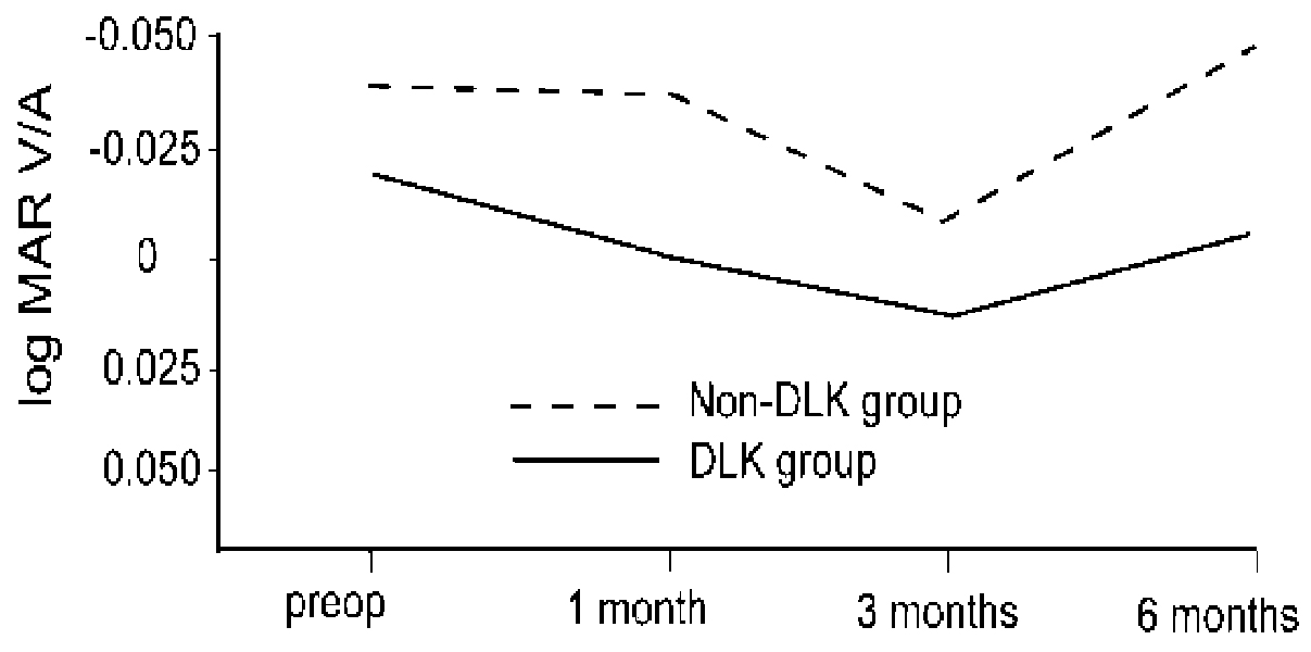
Fig. 1 Comparison of the changes of visual acuity between the DLK and non-DLK groups following LASIK. The visual acuity of the non-DLK group was better at all times, but the difference was not statistically significant (p>0.05). MAR, minimal angle of resolutions; V/A, visual acuity; DLK, diffuse lamellar keratitis; preop, preoperative.

Fig. 2 Comparison of the changes of log contrast sensitivities (CS) three months following LASIK, compared to preoperative level. A: changes of logCS in diffuse lamellar keratitis (DLK) group after three months following LASIK. CS in 12 CPD was significantly decreased at three months postoperatively in the DLK group (*p=0.023, Wilcoxon signed rank test). B: changes of logCS in those without DLK. There were no changes in CS. CS, contrast sensitivity; DLK, diffuse lamellar keratitis; CPD, cycles per degree.
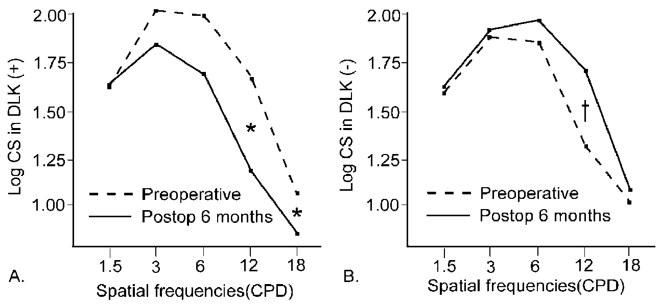
Fig. 3 Comparison of the changes of log contrast sensitivities six months following LASIK, compared to preoperative level. A: changes of log contrast sensitivity (CS) in diffuse lamellar keratitis (DLK) group after six months following LASIK. CS in 12 and 18 CPD were significantly decreased at six months postoperatively in the DLK group (*p=0.043, 0.045, respectively from left, Wilcoxon signed rank test). B: changes of logCS in those without DLK. CS in 12 CPD was significantly increased at six months postoperatively in the non-DLK group (†p=0.042, Wilcoxon signed rank test). CS, contrast sensitivity; DLK, diffuse lamellar keratitis; CPD, cycles per degree.
Reference
-
1. Chan JW, Edwards MH, Woo GC, Woo VC. Contrast sensitivity after laser in situ keratomileusis: one-year follow-up. J Cataract Refract Surg. 2002. 28:1774–1779.2. Nakamura K, Bissen-Miyajima H, Toda I, et al. Effect of laser in situ keratomileusis correction on contrast visual acuity. J Cataract Refract Surg. 2001. 27:357–361.3. Holladay JT, Dudeja DR, Chang J. Functional vision and corneal changes after laser in situ keratomileusis determined by contrast sensitivity, glare testing, and corneal topography. J Refract Surg. 1999. 25:663–669.4. Smith RJ, Maloney RK. Diffuse lamellar keratitis: a new syndrome in lamellar refractive surgery. Ophthalmology. 1998. 105:1721–1726.5. Kaufman SC, Maitchouk DY, Chiou AG, Beuerman RW. Interface inflammation after laser in situ keratomileusis. Sands of the Sahara syndrome. J Cataract Refract Surg. 1998. 24:1589–1593.6. Linebarger EJ, Hardten DR, Lindstrom RL. Diffuse lamellar keratitis: diagnosis and management. J Cataract Refract Surg. 2000. 26:1072–1077.7. Johnson JD, Harissi-Dagher M, Pineda R, et al. Diffuse lamellar keratitis: incidence, associations, outcomes, and a new classification system. J Cataract Refract Surg. 2001. 27:1560–1566.8. Amano R, Ohno K, Shimizu K, et al. Late-onset diffuse lamellar keratitis. Jpn J Ophthalmol. 2003. 47:463–468.9. Belda JI, Artola A, Alio J. Diffuse lamellar keratitis 6 months after uneventful laser in situ keratomileusis. J Refract Surg. 2003. 19:70–71.10. Yuhan KR, Nguyen L, Wachler BS. Role of instrument cleaning and maintenance in the development of diffuse lamellar keratitis. Ophthalmology. 2002. 109:400–404.11. Levinger S, Landau D, Kremer I, et al. Wiping microkeratome blades with sterile 100% alcohol to prevent diffuse lamellar keratitis after laser in situ keratomileusis. J Cataract Refract Surg. 2003. 29:1947–1949.12. Thammano P, Rana AN, Talamo JH. Diffuse lamellar keratitis after laser in situ keratomileusis with the Moria LSK-One and Carriazo-Barraquer microkeratomes. J Cataract Refract Surg. 2003. 29:1962–1968.13. Stulting RD, Randleman JB, Couser JM, Thompson KP. The epidemiology of diffuse lamellar keratitis. Cornea. 2004. 23:680–688.14. Wilson SE, Ambrosio R Jr, Wilson SE, Ambrosio R Jr. Sporadic diffuse lamellar keratitis (DLK) after LASIK. Cornea. 2002. 21:560–563.15. Hoffman RS, Fine IH, Parker M. Incidence and outcomes of LASIK with diffuse lamellar keratitis treated with topical and oral corticosteroids. J Cataract Refract Surg. 2003. 29:451–456.16. Schade OH Sr. Optical and photoelectric analog of the eye. J Opt Soc Am. 1956. 46:721–739.17. Perez-Santonja JJ, Sakla HF, Alio JL. Contrast sensitivity after laser in situ keratomileusis. J Cataract Refract Surg. 1998. 24:183–189.18. Jindra LF, Zemon V. Contrast sensitivity testing: a more complete assessment of vision. J Cataract Refract Surg. 1989. 15:141–148.19. Mutyala S, McDonald MB, Scheinblum KA, et al. Contrast sensitivity evaluation after laser in situ keratomileusis. Ophthalmology. 2000. 107:1864–1867.20. Quesnel NM, Lovasik JV, Ferremi C, et al. Laser in situ keratomileusis for myopia and the contrast sensitivity function. J Refract Surg. 2004. 30:1209–1218.21. Montes-Mico R, Espana E, Menezo JL. Mesopic contrast sensitivity function after laser in situ keratomileusis. J Refract Surg. 2003. 19:353–356.22. Montes_Mico R, Charman WN. Choice of spatial frequency for contrast sensitivity evaluation after corneal refractive surgery. J Refract Surg. 2001. 17:646–651.23. Yamane N, Miyata K, Samejima T, et al. Ocular higher-order aberrations and contrast sensitivity after conventional laser in situ keratomileusis. Invest Ophthalmol Vis Sci. 2004. 45:3986–3990.24. Langrova H, Derse M, Hejcmanova D, et al. Effect of photorefractive keratectomy and laser in situ keratomileusis in high myopia on logMAR visual acuity and contrast sensitivity. Acta Medica (Hradec Kralove). 2003. 46:15–18.25. Moon CS, Tchah HW. Results of LASIK for high myopia. J Korean Ophthalmol Soc. 1998. 39:865–871.26. Cho SW, Kim HM. Comparison of the clinical results of lensectomy and LASIK for high myopia. J Korean Ophthalmol Soc. 1998. 39:1697–1706.27. Vajpayee RB, Balasubramanya R, Rani A, et al. Visual performance after interface haemorrhage during laser in situ keratomileusis. Br J Ophthalmol. 2003. 87:717–719.
- Full Text Links
-
- Actions
-
Cited
- CITED
-
- Close
- Share
-
- Similar articles
-
- Sands of the Sahara Syndrome
- Flap Complications of LASIK
- Comparison of Wave-front Guided LASIK and Conventional LASIK
- A Comparative Study for Mesopic Contrast Sensitivity between Keratectomy(PRK) and Laser in Situ Keratomileusis(LASIK)
- Central Toxic Keratopathy after Femtosecond Laser in-situ Keratomileusis
