Ultrasonographic evaluation of tracheal collapse in dogs
- Affiliations
-
- 1Department of Veterinary Diagnostic Imaging, College of Veterinary Medicine, Konkuk University, Seoul 143-701, Korea.
- 2Department of Veterinary Surgery, College of Veterinary Medicine, Kyungpook National University, Daegu 702-701, Korea. khojang@knu.ac.kr
- 3Department of Veterinary Internal Medicine, College of Veterinary Medicine, Kyungpook National University, Daegu 702-701, Korea.
- 4Department of Veterinary Anatomy, College of Veterinary Medicine, Kyungpook National University, Daegu 702-701, Korea.
- KMID: 1104916
- DOI: http://doi.org/10.4142/jvs.2008.9.4.401
Abstract
- Tracheal ultrasonography was performed to measure the width of the tracheal ring shadow and to assess the clinical relevance of these measurements for identifying tracheal collapse. The first tracheal ring width (FTRW) and thoracic inlet tracheal ring width (TITRW) were measured on both expiration and inspiration. The mean of the FTRW width (129 dogs) was greater in expiration (10.97 +/- 1.02 mm, p = 0.001) than that in inspiration (9.86 +/- 1.03 mm). For 51 normal dogs, the mean of the TITRW width was greater in expiration (9.05 +/- 1.52 mm, p = 0.001) than in inspiration (8.02 +/- 1.43 mm). For 78 tracheal collapse dogs, the mean of the TITRW width was greater in expiration (15.89 +/- 1.01 mm, p = 0.001) than in inspiration (14.85 +/- 1.17 mm). The TITRW/FTRW ratio of the normal dogs was higher (p = 0.001) in expiration (0.81 +/- 0.09) than that in inspiration (0.79 +/- 0.10). When compared between the normal and tracheal collapse dogs, the TITRW/FTRW ratio was also increased (p = 0.001) both in expiration (1.54 +/- 0.09) and inspiration (1.47 +/- 0.08), respectively. Based on these results, the cutoff level of the TITRW/FTRW ratio was statistically analyzed according to the receiver operating characteristic curve and it could be set at 1.16 in expiration and at 1.13 in inspiration. We have demonstrated that tracheal ultrasonography is a useful technique for the evaluation of tracheal collapse and it can be a supportive tool together with the radiographic findings for making the correct diagnosis.
MeSH Terms
Figure
-

Fig. 1 Normal transverse image of the first tracheal ring (A) and the thoracic inlet tracheal ring (B) of a 4-year-old Yorkshire Terrier. An oval shaped hyperechoic tracheal ring (empty arrows) can be seen. The tracheal ring width can be measured between the end points (white arrows) that produce acoustic shadowing. The FTRW and TITRW are 13.4 mm and 11.1 mm, respectively. The sternohyoid (*), sternothyroid (white arrowhead) and sternocephalicus (two arrowheads) muscles are indicated. C: carotid artery, J: jugular vein, Bc: brachiocephalicus muscle, Lc: longus capitis muscle, TG: thyroid gland.
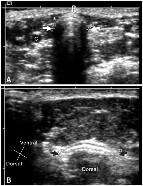
Fig. 2 Transverse image in a 7-year-old Miniature Poodle with severe tracheal collapse. The first tracheal ring (A) shows a semicircular shadow, but the thoracic inlet tracheal ring (B) is flattened and displaced laterally. The sternohyoid muscle (*) and carotid artery (C) are seen. The FTRW (white arrows) and TITRW (black arrows) are 11.3 mm and 17.1 mm, respectively.
Reference
-
1. Buback JL, Boothe HW, Hobson HP. Surgical treatment of tracheal collapse in dogs: 90 cases (1983-1993). J Am Vet Med Assoc. 1996. 208:380–384.2. Dallman MJ, Brown EM. Statistical analysis of selected tracheal measurements in normal dogs and dogs with collapsed trachea. Am J Vet Res. 1984. 45:1033–1037.3. Dabanoğlu I, Öcal MK, Kara ME. A quantitative study on the trachea of the dog. Anat Histol Embryol. 2001. 30:57–59.
Article4. Done SH, Drew RA. Observations on the pathology of tracheal collapse in dogs. J Small Anim Pract. 1976. 17:738–791.
Article5. Ettinger SJ, Kantrowitz B. Ettinger SJ, Feldman EC, editors. Diseases of the trachea. Textbook of Veterinary Internal Medicine: Diseases of the Dog and Cat. 2005. 6th ed. St. Louis: Saunders;1217–1232.6. Fingland RB, Layton CI, Kennedy GA, Galland JC. A comparison of simple continuous versus simple interrupted suture patterns for tracheal anastomosis after large-segment tracheal resection in dogs. Vet Surg. 1995. 24:320–330.
Article7. Harvey CE, Fink EA. Tracheal diameter: Analysis of radiographic measurements in brachycephalic and nonbrachycephalic dogs. J Am Anim Hosp Assoc. 1982. 18:570–576.8. Harvey CE. Review of results of airway obstruction surgery in the dogs. J Small Anim Pract. 1983. 24:555–559.9. Herrtage ME, White R. Bonagura JD, Kirk RW, editors. Management of tracheal collapse. Kirk's Current Veterinary Therapy XIII: Small Animal Practice. 2000. Philadelphia: Saunders;796–801.10. Kealy JK, McAllister H. Diagnostic Radiology and Ultrasonography of the Dog and Cat. 2005. 4th ed. St.Louis: Saunders;173–295.11. Kneller SK. Thrall DE, editor. The Larynx, Pharynx and Trachea. Textbook of Veterinary Diagnostic Radiology. 2002. 4th ed. Philadelphia: Saunders;323–328.12. Nelson AW. Slatter DH, editor. Disease of the trachea. Textbook of Small Animal Surgery. 2003. 3rd ed. Philadelphia: Saunders;858–862.13. Nyland TG, Mattoon JS. Small Animal Diagnostic Ultrasound. 2002. 2nd ed. Philadelphia: Saunders;285–304.14. Rudorf H, Herrtage ME, White RA. Use of ultrasonography in the diagnosis of tracheal collapse. J Small Anim Pract. 1997. 38:513–518.
Article15. Torrez CV, Hunt GB. Results of surgical correction of abnormalities associated with brachycephalic airway obstruction syndrome in dogs in Australia. J Small Anim Pract. 2006. 47:150–154.
Article16. White RAS, William JM. Tracheal collapse in the dog: is there really a role for surgery? A survey of 100 cases. J Small Anim Pract. 1994. 35:191–196.
Article
- Full Text Links
-
- Actions
-
Cited
- CITED
-
- Close
- Share
-
- Similar articles
-
- The Safety and efficacy of a new self-expandable intratracheal nitinol stent for the tracheal collapse in dogs
- Surgical outcomes in dogs with tracheal collapse treated with a novel crossand-hook braided endoluminal stent
- Fluoroscopic characteristics of tracheal collapse and cervical lung herniation in dogs: 222 cases (2012–2015)
- The role of omentopexy in tracheal transplantation in dogs
- Laryngeal Mask Airway as a Conduit for Fiberoptic Intubation and Tracheal Evaluation: 2 Cases report
