Primary chondrosarcoma in the skull of a dog
- Affiliations
-
- 1Department of Veterinary Surgery, Faculty of Agriculture, Yamaguchi University, Yoshida, Yamaguchi 753-8515, Japan. nakaichi@yamaguchi-u.ac.jp
- KMID: 1104243
- DOI: http://doi.org/10.4142/jvs.2007.8.1.99
Abstract
- Chondrosarcoma of the skull is a rare primary malignant tumor that is slow-growing, but locally aggressive. A 5-year-old, golden retriever was presented to our hospital with a swelling in the left side of her head, and the swelling had slowly enlarged over the previous month. There were no significant changes on the neurological examination. A computed tomography scan revealed a large mass involving bone destruction and prominent matrix mineralization. T1-weighted magnetic resonance imaging showed a slightly low-signal intensity area and a T2-weighted image revealed marked, high-signal intensity. There was compression of the adjacent brain parenchyma. Histopathological examination confirmed the lesion to be a chondrosarcoma.
MeSH Terms
Figure
-
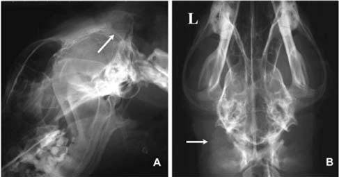
Fig. 1 Radiograph of the head revealed lytic lesion with endosteal scalloping (A, arrow) and calcification within the soft tissue (B, arrow).
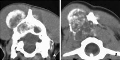
Fig. 2 CT scan showed bone destruction of the skull (A) and matrix calcification (A, B).

Fig. 3 Transverse T1-weighted MRI (A) and its contrastenhanced image (C) showed a slightly low-signal intensity area and mild marginal enhancement around the mass, respectively. T2-weighted image revealed the marked high-signal intensity (B, D) and some compression of the adjacent brain parenchyma (D).
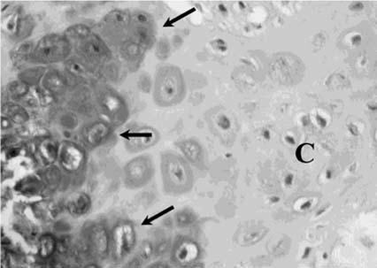
Fig. 4 Histopathological finding revealed proliferation of the atypical chondrocytes (arrows) and cartilaginous component (C). H&E stain, ×400.
Cited by 1 articles
-
Multilobular tumour of the caudal cranium causing severe cerebral and cerebellar compression in a dog
Vassilios Psychas, Panayiotis Loukopoulos, Zoe S. Polizopoulou, Georgios Sofianidis
J Vet Sci. 2009;10(1):81-83. doi: 10.4142/jvs.2009.10.1.81.
Reference
-
1. Burk RL, Ackerman M. Small Animal Radiology and Ultrasonography: a Diagnostic Atlas and Text. 1996. 2nd. Philadelphia: Saunders;427–530.2. Davis GJ, Holt D. Two chondrosarcomas in the urethra of a German shepherd dog. J Small Anim Pract. 2003. 44:169–171.3. Koch BB, Karnell LH, Hoffman HT, Apostolakis LW, Robinson RA, Zhen W, Menck HR. National cancer database report on chondrosarcoma of the head and neck. Head Neck. 2000. 22:408–425.
Article4. Lee YY, Van Tassel P. Craniofacial chondrosarcomas: imaging findings in 15 untreated cases. AJNR Am J Neuroradiol. 1989. 10:165–170.5. Lipsitz D, Levitski RE, Berry WL. Magnetic resonance imaging features of multilobular osteochondrosarcoma in 3 dogs. Vet Radiol Ultrasound. 2001. 42:14–19.
Article6. Littrell LA, Wenger DE, Wold LE, Bertoni F, Unni KK, White LM, Kandel R, Sundaram M. Radiographic, CT, and MR imaging features of dedifferentiated chondrosarcomas: a retrospective review of 174 de novo cases. Radiographics. 2004. 24:1397–1409.
Article7. Murphey MD, Walker EA, Wilson AJ, Kransdorf MJ, Temple HT, Gannon FH. From the archives of the AFIP: imaging of primary chondrosarcoma: radiologic-pathologic correlation. Radiographics. 2003. 23:1245–1278.8. Popvitch CA, Weinstein MJ, Goldschmidt MH, Shofer FS. Chondrosarcoma: a retrospective study of 97 dogs (1987-1990). J Am Anim Hosp Assoc. 1994. 30:81–85.9. Straw RC. Withrow SJ, MacEwen EG, editors. Tumors of the skeletal system. Small Animal Clinical Oncology. 1996. 2nd. Philadelphia: Saunders;287–315.10. Varma DGK, Ayala AG, Carrasco CH, Guo SQ, Kumar R, Edeiken J. Chondrosarcoma: MR imaging with pathologic correlation. Radiographics. 1992. 12:687–704.
Article
- Full Text Links
-
- Actions
-
Cited
- CITED
-
- Close
- Share
-
- Similar articles
-
- A Primary Ossifying Intracranial Myxoma Arising from the Ethmoid Sinus
- Chondrosarcoma of the Parasella Area: Case Report
- Chondrosarcoma with Intratumoral Hemorrhage: Case Report
- Chondrosarcoma of the Skull Base ; Report of Three Cases
- Mesenchymal chondrosarcoma in the maxillary gingiva of a Maltese dog: a case report
