J Vet Sci.
2009 Jun;10(2):173-175. 10.4142/jvs.2009.10.2.173.
Dilation of the olfactory bulb cavity concurrent with hydrocephalus in four small breed dogs
- Affiliations
-
- 1BK21 Basic & Diagnostic Veterinary Specialist Program for Animal Diseases and Department of Veterinary Internal Medicine, College of Veterinary Medicine, Konkuk University, Seoul 143-701, Korea. parkhee@konkuk.ac.kr
- 2College of Electronics and Information, Kyunghee University, Yongin 446-701, Korea.
- KMID: 1102977
- DOI: http://doi.org/10.4142/jvs.2009.10.2.173
Abstract
- Four small breed dogs were admitted with seizures. Magnetic resonance imaging (MRI) of the brain revealed dilation of the olfactory bulb cavity as well as enlargement of the lateral ventricles. These findings demonstrate that dilation of the olfactory bulb cavity can occur concurrent with hydrocephalus. This is the first description of the clinical and MRI features of dilation of the olfactory bulb cavity concurrent with hydrocephalus in dogs.
Keyword
MeSH Terms
Figure
-
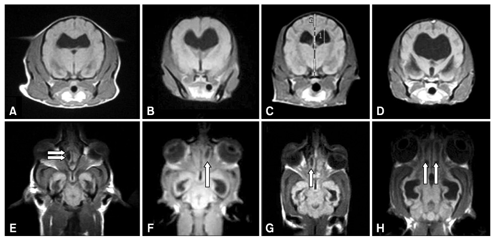
Fig. 1 MR images obtained from patients. (A-D) Transverse T1-weighted images (WI) of Case 1, 2, 3, and 4 show the enlarged lateral ventricles, respectively. (E-H) Dorsal T1-WI of Case 1, 2, 3, and 4 show hypointensity lesion (arrows) in olfactory bulb, respectively.
Reference
-
1. Brinker T, Lüdemann W, Berens von Rautenfeld D, Samii M. Dynamic properties of lymphatic pathways for the absorption of cerebrospinal fluid. Acta Neuropathol. 1997. 94:493–498.
Article2. Dewey CW. A Practical Guide to Canine and Feline Neurology. 2003. 1st ed. Ames: Wiley-Blackwell;36107–110.3. Friedman DI. Pseudotumor cerebri. Neurol Clin. 2004. 22:99–131.
Article4. Johnston M, Zakharov A, Papaiconomou C, Salmasi G, Armstrong D. Evidence of connections between cerebrospinal fluid and nasal lymphatic vessels in humans, non-human primates and other mammalian species. Cerebrospinal Fluid Res. 2004. 1:2.5. Kapoor KG, Katz SE, Grzybowski DM, Lubow M. Cerebrospinal fluid outflow: an evolving perspective. Brain Res Bull. 2008. 16:327–334.
Article6. Lorenz MD, Kornegay JN. Handbook of Veterinary Neurology. 2004. 4th ed. Philadelphia: Saunders;313–318.7. Mollanji R, Papaiconomou C, Boulton M, Midha R, Johnston M. Comparison of cerebrospinal fluid transport in fetal and adult sheep. Am J Physiol Regul Integr Comp Physiol. 2001. 281:R1215–R1223.
Article8. Rammling M, Madan M, Paul L, Behnam B, Pattisapu JV. Evidence for reduced lymphatic CSF absorption in the H-Tx rat hydrocephalus model. Cerebrospinal Fluid Res. 2008. 5:15.
Article9. Selby LA, Hayes HM Jr, Becker SV. Epizootiologic features of canine hydrocephalus. Am J Vet Res. 1979. 40:411–413.10. Smith BJ. Canine Anatomy. 1999. 1st ed. Philadelphia: Lippincott Williams & Wilkins;152–161.11. Weller RO, Kida S, Zhang ET. Pathways of fluid drainage from the brain--morphological aspects and immunological significance in rat and man. Brain Pathol. 1992. 2:277–284.
Article
- Full Text Links
-
- Actions
-
Cited
- CITED
-
- Close
- Share
-
- Similar articles
-
- Magnetic resonance imaging features of syringobulbia in small breed dogs
- Gait analysis in clinically healthy small to toy breed dogs using a pressure plate
- Anatomical atudy of the olfactory bulb in the cat
- Structural changes of the synapses within glomeruli of the olfactory bulb after lesion of olfactory epithelium in the rat
- MR Evaluation of the Olfactory Bulb in Normals and Patients with Decreased Sense of Smell*
