Two different types of malignant fibrous histiocytomas from pet dogs
- Affiliations
-
- 1Department of Clinical Pathology, College of Veterinary Medicine, Konkuk University, Seoul 143-701, Korea.
- 2Department of Pathology, College of Veterinary Medicine, Kyungpook National University, Daegu 702-701, Korea. jeongks@knu.ac.kr
- KMID: 1102976
- DOI: http://doi.org/10.4142/jvs.2009.10.2.169
Abstract
- We describe 2 cases of malignant fibrous histiocytomas (MFHs) that spontaneously developed in young pet dogs. To classify these tumors, we applied a panel of antibodies (vimentin, desmin, alpha-SMA, and ED1) and Azan staining for collagen. The MFHs were most consistent with osteoclast-like giant and inflammatory cell types. The first case had positive staining for ED1 and vimentin, and given the osteoclast-like giant cells, calcification sites accompanying peripheral giant cell infiltrates. The latter case, the inflammatory cell type, exhibited a storiform-pleomorphic variant of neoplastic cells, including an ossifying matrix. MFHs are among the most highly aggressive tumors occurring in soft tissue sarcomas in elderly dogs; however, MFHs have been poorly studied from a diagnostic point of view. Herein, we describe the histologic and immunohistologic features of MFHs in detail, thus classifying the subtypes of these tumors.
MeSH Terms
Figure
-
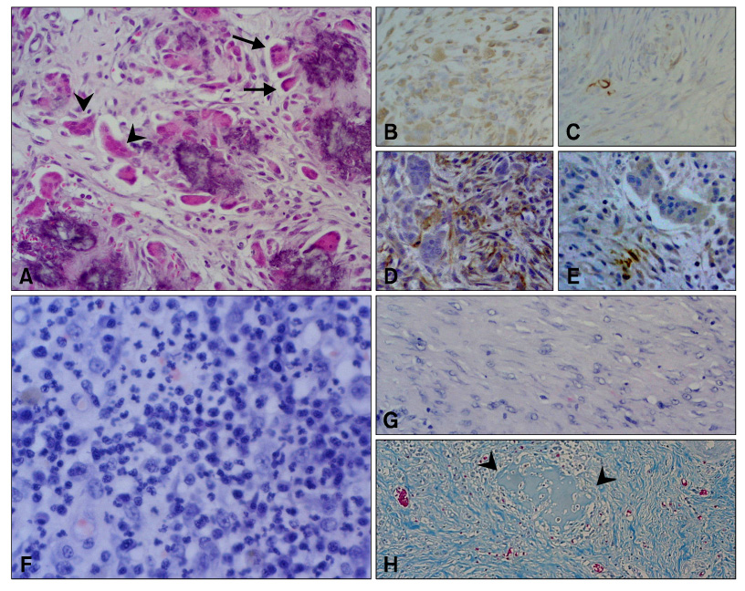
Fig. 1 Malignant fibrous histiocytoma (MFH), subtype osteoclast-like giant cell type (A-E). Osteoclast-like giant cells (arrow head) resembling normal osteoclast are scattered throughout the lesion, especially around the calcification sites. In addition to these features, peripheral giant cells (arrow) appeared (A). Immunostaining for vimentin, desmin, α-SMA, and ED1, respectively (B-E). Tumor cells were positive for vimentin (B), and negative for α-SMA (D) and ED1 (E). Note the strong positive for desmin of smooth muscle cells of blood vessels vs. negative staining of tumor cells (C). Osteoclast-like giant cells showed positive for ED1 (E). Inflammatory cell type of MFH (F-H). This type of MHF was characterized by infiltration of various inflammatory cells (F) and ossifying matrix (G). Note the bone matrix surrounding the neoplastic fibroblasts (H). A, F and G: H&E stain, B, C, D and E: ABC method counterstained with hematoxylin, H: Azan stain. A-E: ×66, F and G: ×132, H: ×33.
Reference
-
1. Carip C, de Beaumont T. Malignant histiocytofibroma of the small intestine in a young immune deficient patient. Presse Med. 2002. 31:214–216.2. Folpe AL, Morris RJ, Weiss SW. Soft tissue giant cell tumor of low malignant potential: a proposal for the reclassification of malignant giant cell tumor of soft parts. Mod Pathol. 1999. 12:894–902.3. Gregory M, Barbara EP, Dennis M, Stephen JW. Small Animal Clinical Oncology. 2001. 3rd ed. Pennsylvenia: Saunders;283–287.4. Lew W, Lim HS, Kim YC. Cutaneous metastatic malignant fibrous histiocytoma. J Am Acad Dermatol. 2003. 48:S39–S40.
Article5. Morris JS, McInnes EF, Bostock DE, Hoather TM, Dobson JM. Immunohistochemical and histopathologic features of 14 malignant fibrous histiocytomas from Flat-Coated Retrievers. Vet Pathol. 2002. 39:473–479.
Article6. Orlandi A, Bianchi L, Ferlosio A, Innocenzi I, Spagnoli LG. The origin of osteoclast-like giant cells in atypical fibroxanthoma. Histopathology. 2003. 42:407–410.
Article7. Pérez-Martínez C, García Fernnádez RA, Reyes Avila LE, Pérez-Pérez V, González N, García-Iglesias MJ. Malignant fibrous histiocytoma (giant cell type) associated with a malignant mixed tumor in the salivary gland of a dog. Vet Pathol. 2000. 37:350–353.
Article8. Rebecca B, John HL. Color Atlas of Cytology of the Dog and Cat. 2000. Missouri: Mosby;46.9. Kaddu S, McMenamin ME, Fletcher CD. Atypical fibrous histiocytoma of the skin: clinicopathologic analysis of 59 cases with evidence of infrequent metastasis. Am J Surg Pathol. 2002. 26:35–46.10. Wiriosuparto S, Krassilnik N, Gologan A, Cohen JM, Wenig B. Malignant fibrous histiocytoma, giant cell type, of the breast mimicking metaplastic carcinoma. A case report. Acta Cytol. 2003. 47:673–678.
Article
- Full Text Links
-
- Actions
-
Cited
- CITED
-
- Close
- Share
-
- Similar articles
-
- Ultrastructure of 2 Malignant Fibrous Histiocytomas with Reference to the Histogenesis
- ras Gene Mutations in Malignant Fibrous Histiocytoma
- A Case of Malignant Fibrous Histiocytoma in the Conjunctiva
- Expression of Matrix Metalloproteinase-9 Correlates with Poor Prognosis in Human Malignant Fibrous Histiocytoma
- Malignant Fibrous Histiocytoma of the Stomach - A case repot -
