Lesions in the thymus and bone marrow in chicks with experimentally induced chicken infectious anemia disease
- Affiliations
-
- 1Department of Pathology, Faculty of Veterinary Medicine, Istanbul University, Avcilar 34310, Istanbul, Turkey. Agurel@istanbul.edu.tr
- KMID: 1102949
- DOI: http://doi.org/10.4142/jvs.2008.9.1.15
Abstract
- One-day-old SPF chicks were inoculated with the Cux-l strain of chicken infectious anemia virus (CIAV), and the clinical development of disease and its macroscopic and microscopic alterations in the thymus and bone marrow, were observed. Tissue sections of thymus and bone marrow were stained using the streptavidin-biotin peroxidase method and examined under light microscope for evaluation of antigenic intensities in tissues. Those findings were then compared with blood parameters and ELISA results obtained through collected sera during sacrifice procedures. We sought to determine: the localization of viral antigens in thymus and bone marrow tissues after inoculation, the correlation between antigen intensities and hematologic, serologic and histopathologic findings, definitive diagnostic criteria using histopathologic and immunoperoxidase methods, and the reliability of these methods in the diagnosis of CIAV infection. For this purpose, 83, one-day-old SPF chicks were used. The birds were divided into experimental (n = 52) and control (n = 26) groups. A virus dose of TCID50 of 100,000/ml was administered intramuscularly to every bird in the experimental group. Based on the results of this study, we have suggested that clinical examination, along with macroscopic and microscopic evaluation of the thymus and bone marrow, maybe undertaken starting from day 7 post-inoculation (PI). ELISA, might be of value, as it might give consistent results starting from day 14 PI. However, the most reliable results were obtained through examination of thymus and bone marrow sections from infected birds stained by immunoperoxidase technique, as early as day 4 PI.
MeSH Terms
Figure
-

Fig. 1 (A) Normal appearance of the thymus of a bird from the control group sacrificed on day 12. Scale bar = 200 µm. (B) Depletion of the cortical thymocytes and medulla-like appearance of the thymic cortex in the thymus section of a bird from the experimental group sacrificed on day 12. Scale bar = 200 µm. (C) Normal appearance of the bone marrow of a bird from the control group sacrificed on day 10. Scale bar = 100 µm. (D) A prominent decrease in the number of cells in the bone marrow of a bird from the experimental group sacrificed on day 10. Scale bar = 100 µm. H&E stain.
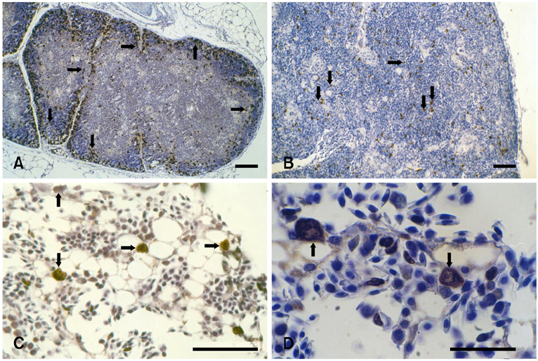
Fig. 2 (A) Abundant antigen-specific staining in the cortex and, to a lesser extent, in the medulla of the thymus section of an experimental bird sacrificed on day 10. Scale bar = 100 µm. (B) Antigen-specifically stained cells both in the depleted cortical and medullar zones of the thymus section of an experimental bird sacrificed on day 12. Scale bar = 200 µm. (C) Antigen-specifically stained large hemocytoblasts in the bone marrow section of an experimental bird sacrificed on day 8. Scale bar = 100 µm. (D) Antigen-specifically stained hemocytoblasts and stem cells at different stages of differentiation in the bone marrow section of an experimental bird sacrificed on day 10. Scale bar = 50 µm. Immunoperoxidase stain.
Reference
-
1. Bülow VV. Avian infectious anemia and related syndromes caused by chicken anemia virus. Crit Rev Poult Biol. 1991. 3:1–17.2. Bülow VV, Sebat KA. Calnek BW, Barnes HJ, Beard CW, McDonald LR, Saif YM, editors. Chicken infectious anemia. Disease of Poultry. 1997. 10th ed. Ames: Iowa State University Press;739–756.3. Duncan DB. Multiple range and multiple F tests. Biometrics. 1955. 11:1–42.
Article4. Ergün A, Yurtman M, Nalbantsoy A. A sero-survey study on chicken anemia (CAV) by ELISA test. Ciftlik. 1998. 171:44–46.5. Goodwin MA, Brown J. Inability of so-called chicken anemia agent (CAA) infections to be diagnosed by anemia and hematopoietic organ atrophy alone. Avian Dis. 1992. 36:353–355.
Article6. Goodwin MA, Brown J, Davis JF, Girshick T, Miller SL, Nordgren RM, Rodenberg J. Comparisons of packed cell volumes (PCVs) from so-called chicken anemia agent (CAA; a virus)-free broilers to PCVs from CAA-free specific-pathogen-free leghorns. Avian Dis. 1992. 36:1063–1066.
Article7. Goodwin MA, Brown J, Latimer KS, Miller SL. Packed cell volume reference intervals to aid in the diagnosis of anemia and polycythemia in young leghorn chickens. Avian Dis. 1991. 35:820–823.
Article8. Goodwin MA, Davis JF, Brown J. Packed cell volume reference intervals to aid in the diagnosis of anemia and polycythemia in young broiler chickens. Avian Dis. 1992. 36:440–443.
Article9. Goodwin MA, Smeltzer MA, Brown J, Girshick T, McMurray BL, McCarter S. Effect of so-called chicken anemia agent maternal antibody on chick serologic conversion to viruses in the field. Avian Dis. 1993. 37:542–545.
Article10. Goryo M, Suwa T, Umemura T, Itakura C, Yamashiro S. Histopathology of chicks inoculated with chicken anemia agent (MSB1-TK5803 strain). Avian Pathol. 1989. 18:73–89.
Article11. Gown AM, de Wever N, Battifora H. Microwave-based antigenic unmasking. A revolutionary new technique for routine immunohistochemistry. Appl Immunohistochem. 1993. 1:256–266.12. Hoop RK. Persistence and vertical transmission of chicken anemia agent in experimentally infected laying hens. Avian Pathol. 1992. 21:493–501.
Article13. Hoop RK, Reece RL. The use of immunofluorescence and immunoperoxidase staining in studying the pathogenesis of chicken anemia agent in experimentally infected chickens. Avian Pathol. 1991. 20:349–355.
Article14. Inoue M, Fukuda M, Miyano K. Thymic lesions in chicken infected with infectious bursal disease virus. Avian Dis. 1994. 38:839–846.
Article15. Jeurissen SH, Pol JM, de Boer GF. Transient depletion of cortical thymocytes induced by chicken anaemia agent. Thymus. 1989. 14:115–123.16. McIlroy SG, McNulty MS, Bruce DW, Smyth JA, Goodall EA, Alcorn MJ. Economic effects of clinical chicken anemia agent infection on profitable broiler production. Avian Dis. 1992. 36:566–574.
Article17. McNeilly F, Allan GM, Moffett DA, McNulty MS. Detection of chicken anemia agent in chickens by immunofluorescence and immunoperoxidase staining. Avian Pathol. 1991. 20:125–132.
Article18. McNulty MS. Chicken anemia agent: a review. Avian Pathol. 1991. 20:187–203.19. Michalski WP, O'Rourke D, Bagust TJ. Chicken anemia virus antibody ELISA: problems with non specific reactions. Avian Pathol. 1996. 25:245–254.
Article20. Naish S. Handbook of immunochemical staining methods. 1989. California: Dako;16–20.21. Noteborn MHM, Koch G. Chicken anemia virus infection: molecular basis of pathogenicity. Avian Pathol. 1995. 24:11–13.22. Otaki Y, Saito K, Tajima M, Nomura Y. Persistence of maternal antibody to chicken anemia agent and its effect on the susceptibility of young chickens. Avian Pathol. 1992. 21:147–151.
Article23. Pope CR. Chicken anemia agent. Vet Immunol Immunopathol. 1991. 30:51–65.
Article24. Shi SR, Key ME, Kalra K. Antigen retrieval in formalin-fixed, paraffin-embedded tissues: an enhancement method for immunohistochemical staining based on microwave oven heating of tissue sections. J Histochem Cytochem. 1991. 39:741–748.
Article25. Smyth JA, Moffett DA, McNulty MS, Todd D, Mackie DP. A sequential histopathologic and immunocytochemical study of chicken anemia virus infection at one day of age. Avian Dis. 1993. 37:324–338.
Article26. Taylor CR. Immunoperoxidase techniques. Practical and theoretical aspects. Arch Pathol Lab Med. 1978. 102:113–112.27. Yilmaz H, Turan N, Özgür Y, Helps CR, Akay Ö. Detection of chicken anemia virus DNA in thymus of naturally Infected chicken in Turkey. Avian Dis. 2001. 45:529–533.
Article28. Yuasa N, Taniguchi T, Yoshida I. Isolation and some characteristics of an agent inducing anemia in chicks. Avian Pathol. 1979. 23:366–385.
Article
- Full Text Links
-
- Actions
-
Cited
- CITED
-
- Close
- Share
-
- Similar articles
-
- Bone Marrow Cell Culture(GM-CFU) in Anaplastic Anemia of Children
- Treatment of severe aplastic anemia: comparison between bone marrow transplantation and immunomodulation
- A Case of Ethosuximide-Induced Aplastic Anemia Successfully Treated with Methylprednisolone Pulse Therapy
- Revisiting anemia afer ABO-mismatched allogeneic bone marrow transplantation
- Two Cases of Aplastic Anemia Following Propylthiouracil
