Bilateral Spontaneous Anterior Lens Dislocation in a Retinitis Pigmentosa Patient
- Affiliations
-
- 1Department of Ophthalmology, Kim's Eye Hospital, Myung-Gok Eye Research Institute, Konyang University College of Medicine, Seoul, Korea. yongho@konyang.ac.kr
- KMID: 1101916
- DOI: http://doi.org/10.3341/kjo.2007.21.2.124
Abstract
- PURPOSE: To report a case of bilateral spontaneous anterior lens dislocation associated with retinitis pigmentosa (RP). METHODS: A 45-year-old male with RP presented with elevated intraocular pressure (IOP) in the right eye and was treated with laser iridotomy (LI). After LI, complete crystalline lens dislocation into the anterior chamber occurred. Surgical intervention, including anterior vitrectomy, intracapsular cataract extraction (ICCE), and IOL scleral fixation was performed. Two years later, the same episode occurred in his left eye and a similar treatment was done. RESULTS: Surgery was successful in both eyes. CONCLUSIONS: This is the first report of bilateral spontaneous anterior lens dislocation in a RP patient.
MeSH Terms
-
*Anterior Chamber
Cataract/complications/diagnosis
Cataract Extraction
Electroretinography
Follow-Up Studies
Humans
Iris/surgery
Laser Therapy/adverse effects
Lens Implantation, Intraocular/methods
Lens Subluxation/diagnosis/*etiology/surgery
Male
Middle Aged
Ocular Hypertension/complications/physiopathology/surgery
Retinitis Pigmentosa/*complications/diagnosis/surgery
Sclera/surgery
Suture Techniques
Visual Fields
Vitrectomy
Figure
-
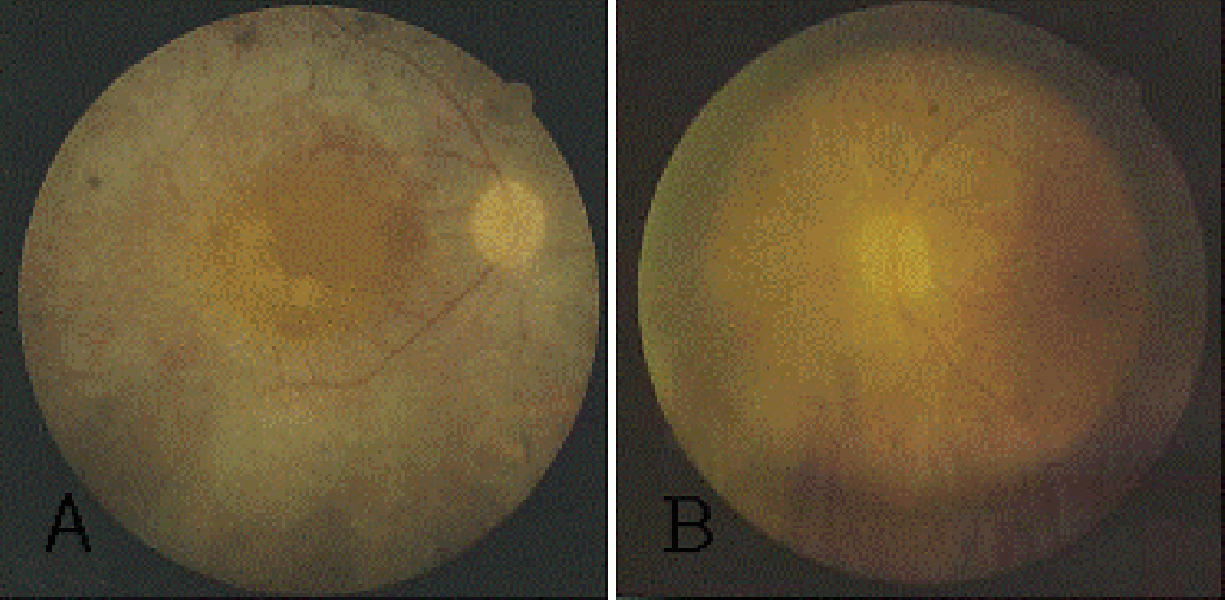
Fig. 1 Fundus photograph of the right (A) and left (B) eye. Bone-spicule pigmentation in the entire retina with sparing of the macula and pale optic discs were noted.

Fig. 2 Visual field of the right (A) and left (B) eye. Extensive peripheral field defect is noted (central 10°). Visual field using the Humphrey Visual Field Analyzer (model 750, Humphrey Instruments, Inc, Dublin, California; C24-2 program).
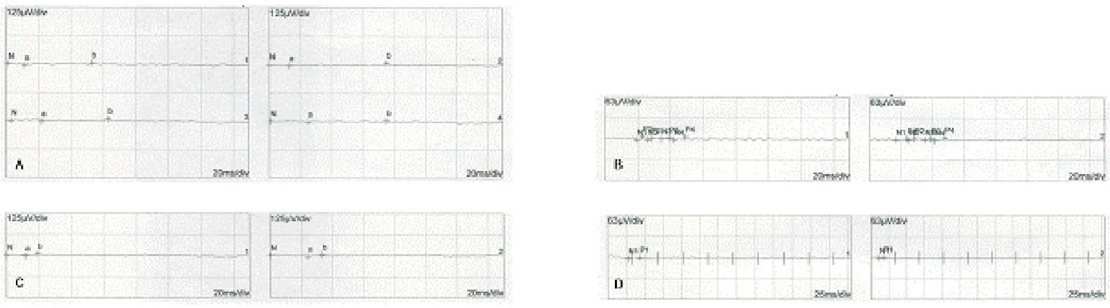
Fig. 3 (A) Scotopic ERG shows prolonged rod a-wave implicit times. (B) Oscillatory ERG. (C) Photopic ERG shows prolonged cone b-wave implicit times. (D) 30Hz Flicker ERG.

Fig. 4 Slit lamp photograph. (A) The preoperative photograph in the left eye with complete anterior lens dislocation. (B) The postoperative photograph in the left eye; dislocated lens was removed and posterior chamber IOL is in place.
Reference
-
1. Ryan S. Retina. 2001. Vol. 1:3rd ed. St, Loius, Missouri: Mosby;362–460.2. Berson EL, Rosner B, Sandberg MA, Dryja TP. Ocular findings in patients with autosomal dominant retinitis pigmentosa and rhodopsin, proline-347-leucine. Am J Ophthalmol. 1991. 111:614–623.3. Sato H, Wada Y, Abe Y, et al. Retinitis Pigmentosa Associated With Ectopia Lentis. Arch Ophthalmol. 2005. 120:852–854.4. Nelson LA, Maumenee IH. Ectopis lentis. Surv Ophthal. 1982. 27:143–160.5. Cross HE, Jensen AD. Ocular manifestations in the Marfan's syndrome and homocystenuria. Am J Ophthalmol. 1973. 75:405–420.6. Hayashi K, Hayashi H, Matsuo K, et al. Anterior capsule contraction and intraocular lens dislocation after implant surgery in eyes with retinitis pigmentosa. Ophthalmology. 1998. 105:1239–1243.7. Namiki M, Tagami Y, Morino I. Findings from slit lamp and historical examination of the anterior capsule in patients with severe anterior capsule shrinkage and opacities after implantation of intraocular lens. J Jpn Ophthalmol Soc. 1993. 97:716–720.8. Allingham R, Damji K, Freedman S, et al. Shields' Textbook of Glaucoma. 2005. 5th ed. Philadelphia: Lippincott Williams & Wilkins;318–327.9. Madill S, Bain K, Patton N, et al. Emergency use of pilocarpine and pupil block glaucoma in ectopia lentis. Eye. 2005. 9:105–107.10. Choi D, Kim J, Song B. Surgical management of crystalline lens dislocation into the anterior chamber with corneal touch and secondary glaucoma. J Cataract Refract Surg. 2004. 30:718–721.11. Young A, Leung G, Chen L, et al. A modified technique of scleral fixated intraocular lenses for aphakic correction. Eye. 2005. 19:19–22.
- Full Text Links
-
- Actions
-
Cited
- CITED
-
- Close
- Share
-
- Similar articles
-
- Bilateral Spontaneous Dislocation of Intraocular Lenses within the Capsular Bag in a Retinitis Pigmentosa Patient
- Visual Function and Functional Vision of Retinitis Pigmentosa
- A Case of Unilateral Retinitis Pigmentosa
- A Case of Retinitis Pigmentosa without Pigment
- Incidence Rate and Risk Factors of Intraocular Lens Dislocation in South Korea
