Anterior Lens Capsule Abnormalities in Alport Syndrome
- Affiliations
-
- 1Department of Ophthalmology, College of Medicine, Konyang University, Myung Gok Eye Research Institute, Daejeon, Korea. eyedr00@yahoo.co.kr
- KMID: 1099092
- DOI: http://doi.org/10.3341/kjo.2005.19.1.84
Abstract
- Alport syndrome is a hereditary, progressive disease characterized by progressive nephritis, sensorineural deafness, and ocular abnormalities, including anterior lenticonus. The ultrastructure of the lens capsule abnormalities in Alport syndrome is reported. Four anterior lens capsules from 31-year-old patient and 26-year-old patient with lenticonus who were affected by the Alport syndrome were obtained at capsulectomy. And all four anterior lens capsules were examined by transmission electron microscopy. The histopathologic findings showed that the thickness of the anterior lens capsules was decreased (4~13 micrometer) and that there were many vascular dehiscences localized at the inner part of the lens capsule. There were large numbers of capsular dehiscences containing fibrillar materials and vacuoles. The anterior capsules were clearly fragile in this disease, forming the basis for the progressive lenticonus and anterior polar cataract.
Keyword
MeSH Terms
Figure
-
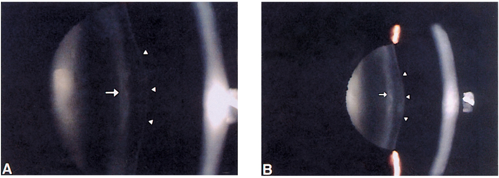
Fig. 1 Slit-lamp photograph showing anterior lenticonus (arrowhead) and anterior subcapsular faint opacity (white arrow). Right eye (A), Left eye (B)

Fig. 2 Fundus photograph in the right eye. Yellow punctate lesions (black arrow) in the retinal pigment epithelium level that spared the macula.

Fig. 3 Transmission electron microscope of anterior lens capsule and epithelium. Capsule (C) is 4.8 µm thick with vertical dehiscences (black arrow). The dehiscences are located in the inner two-thirds of the anterior lens capsule. The outer one-third of the capsule was looser and of fibrillar texture. The lens epithelium (E) is highly irregular shaped. (Original magnification ×5400).

Fig. 4 Transmission electron microscope of the anterior lens capsule. The dehiscences forms a network (black arrow) within the capsule. (Original magnification ×6300).

Fig. 5 High transmission electron microscopic magnification of the anterior lens capsule. Dehiscences contain fibrillar material and vacuole (arrowhead). (Original magnification ×6300).

Fig. 6 Transmission electron microscopic view of the lens epithelium. (A) The lens epithelial cells are highly irregular and their lateral borders are indistinct. The nucleus (N) is smaller, darker, and irregular margined. The cytoplasm contains numerous lacunae (arrowhead). There is a paucity of other organelles. (B) Double-layered lens epithelial cells (black arrow). (C) In focal areas the lens epithelial cells are well preserved. Epithelial cells are cuboidal-shaped and their lateral borders are closed joined.
Cited by 1 articles
-
Bilateral Serous Retinal Detachment Associated With Alport's Syndrome
Young Bin Song, Sung Pyo Park
J Korean Ophthalmol Soc. 2010;51(3):463-468. doi: 10.3341/jkos.2010.51.3.463.
Reference
-
1. Colville D, Savige J. Alport syndrome. A review of the ocular manifestations. Ophthalmic Genet. 1997. 18:161–173.2. Faggioni R, Scouras J, Streiff EB. Alport's syndrome: Clinicopathological considerations. Ophthalmologica. 1972. 165:1–14.3. Gubler M, Levy M, Broyer M, et al. Alport's syndrome: a report of 58 cases and a review of the literature. Am J Med. 1981. 70:493–505.4. Nielsen CE. Lenticonus anterior and Alport's disease. Am J Ophthalmol. 1977. 84:532–535.5. Govan JA. Ocular manifestations of Alport's syndrome : a hereditary disorder of basement membrane. Br J Ophthalmol. 1983. 67:493–503.6. Yoshikawa N, White RH, Cameron AH. Familial hematuria: clinicopathological correlations. Clin Nephrol. 1982. 17:172–182.7. Mayers JC, Jones TA, Pohjolainen ER, et al. Molecular cloning of α5(IV) collagen and assignment of the gene to the region of the X chromosome containing the Alport syndrome locus. Am J Hum Genet. 1990. 46:1024–1033.8. Barker DF, Hostikka SL, Zhou J, et al. Identification of mutations in the COL4A5 collagen gene in Alport syndrome. Science. 1990. 248:1224–1227.9. Mariyama M, Kalluri R, Hudson BG, Reeders ST. The α4(IV) chain of basement membrane collagen : isolation of cDNA encoding bovine α4(IV) and comparison with other type IV collagen. J Biol Chem. 1991. 267:1253–1258.10. Zhou J, Barker DF, Hostikka SL, et al. Single base mutation in α5(IV)collagen chain gene in Alport syndrome. Genomics. 1991. 9:10–18.11. Morrison KE, Germino GG, Reeders ST. Use of the polymerase chain reaction to clone and sequence a cDNA encoding the bovine α3 chain of type IV collagen. J Biol Chem. 1991. 226:34–39.12. Peterson WS, Albert DM. Fundus changes in the hereditary nephropathies. Trans Am Acad Ophthalmol Otolaryngol. 1974. 78:762–771.13. Polak BC, Hogewind BL. Macular lesions in Alport's syndrome. Acta Ophthalmol. 1978. 56:518–530.14. Gupta V, Kumar N. Bilateral macular holes : an unusual feature of Alport syndrome. Retina. 2002. 22:499–501.15. Brownell RD, Wolter JR. Anterior lenticonus in familiar hemorrhagic Nephritis: demonstration of lens pathology. Arch Ophthalmol. 1964. 71:481–483.16. Kato T, Watanabe Y, Nakayasu K, et al. The ultrastructure of the lens capsule abnormalities in Alport's syndrome. Jpn J Ophthalmol. 1998. 42:401–405.17. Ito S, Hataya H, Ikeda M, et al. Alport syndrome-like basement membrane changes in Frasier syndrome: an electron microscopy study. Am J Kidney Dis. 2003. 41:1110–1115.18. Hinglais N, Grunfeld JP, Troconis L, Bois E. Alport's syndrome (progressive hereditary nephritis). Clin Nephrol. 1974. 2:143–156.19. Zyberman R, Silerstone BZ, Brandes E, Drukker A. Retinal lesions in Alport's syndrome. J Pediatr Ophthalmol Strabismus. 1980. 17:255–260.20. Khalil M, Saheb N. Posterior lenticonus. Ophthalmology. 1984. 91:1429–1430.21. Junk AK, Stefani FH, Ludwig K. Bilateral anterior lenticonus: Scheimpflug imaging system documentation and ultrastructural confirmation of Alport syndrome in the lens capsule. Arch Ophthalmol. 2000. 118:895–897.22. Takei K, Furuya A, Hommura S, Yamaugchi N. Ultrastructural fragility and type IV collagen abnormality of the anterior lens capsules in a patient with Alport syndrome. Jpn J Ophthalmol. 2001. 45:103–104.23. Ohkubo S, Takeda H, Higashide T, et al. Immunohistochemical and molecular genetic evidence for type IV collagen a5 chain abnormality in the anterior lenticonus associated with Alport syndrome. Arch Ophthalmol. 2003. 121:846–850.24. Gross O, Netzer KO, Lambrecht R, et al. Novel COL4A4 splice defect and in-frame deletion in a large consanguine family as a genetic link between benign familial hematuria and autosomal Alport syndrome. Nephrol Dial Transplant. 2003. 18:1122–1127.25. Barnard K, Burgers SA, Carter DA. Three-dimensional structure of type IV collagen in the mammalian lens capsule. J Struc Biol. 1992. 108:6–13.26. Wieslander J, Langeveld J, Butkowski R. Physical and immunochemical studies of the globular domain of type IV collagen. Cryptoproperties of the Goodpasture antigen. J Biol Chem. 1985. 260:8564–8570.27. McCoy RC, Johnson HK, Stone WJ, Wilson CB. Absence of nephritogenic GBM antigen(s) in some patients with hereditary nephritis. Kidney Int. 1982. 21:642–652.28. Prakash S, Chung KW, Sinha S, et al. Autosomal dominant progressive nephropathy with deafness: linkage to a new locus on chromosome 11q24. J Am Soc Nephrol. 2003. 14:1794–1803.29. Streeten BW, Robinson MR, Wallace R, Jones DB. Lens capsule abnormalities in Alport's syndrome. Arch Ophthalmol. 1987. 105:1693–1697.
- Full Text Links
-
- Actions
-
Cited
- CITED
-
- Close
- Share
-
- Similar articles
-
- A Case of Anterior Lenticonus in Alport's Syndrome
- Alport Syndrome with Progressive Decrease in Retinal Thickness: A Case Report
- The Capsular Transplantation in Experimentally Induced Capsule Rupture of Porcine Lens
- True Exfoliation of the Lens Capsule
- A Case of Thick Anterior Capsule of Hypermature Cataract in Down Syndrome
