Surgical Management of Bilateral Exudative Retinal Detachment associated with Central Serous Chorioretinopathy
- Affiliations
-
- 1Department of Ophthalmology, Mokdong Hospital, Ewha Womans University College of Medicine, Seoul, Korea. leejhoph@mm.ewha. ac.kr
- 2Department of Ophthalmology, Kangnam Sacred Heart Hospital, Hallym University College of Medicine, Seoul, Korea.
- KMID: 1099050
- DOI: http://doi.org/10.3341/kjo.2006.20.2.131
Abstract
- PURPOSE: To report a case of bilateral bullous exudative retinal detachment in central serous chorioretinopathy (CSC) which was attached by vitrectomy and internal drainage of the subretinal fluid. METHODS: A 47-year-old man affected by bilateral atypical CSC with a bullous retinal detachment with subretinal exudate. A fluorescein angiogram (FAG) showed multiple points of leakage and staining of subretinal fibrosis. A tentative diagnosis of Vogt-Koyanagi-Harada (VKH) syndrome was made and the patient was treated with systemic corticosteroids and immunosuppressive agents. However, the subretinal fluid was not absorbed. He was then treated with vitrectomy and internal drainage of subretinal fluid. RESULTS: The retina was attached successfully in both eyes. Visual acuity improved to 20/50 in his left eye but did not improve in the right eye due to subretinal fibrotic scarring and atropic changes on the macula. CONCLUSIONS: Our case suggests that the surgical management of bullous exudative retinal detachment is safe and necessary.
Keyword
MeSH Terms
Figure
-
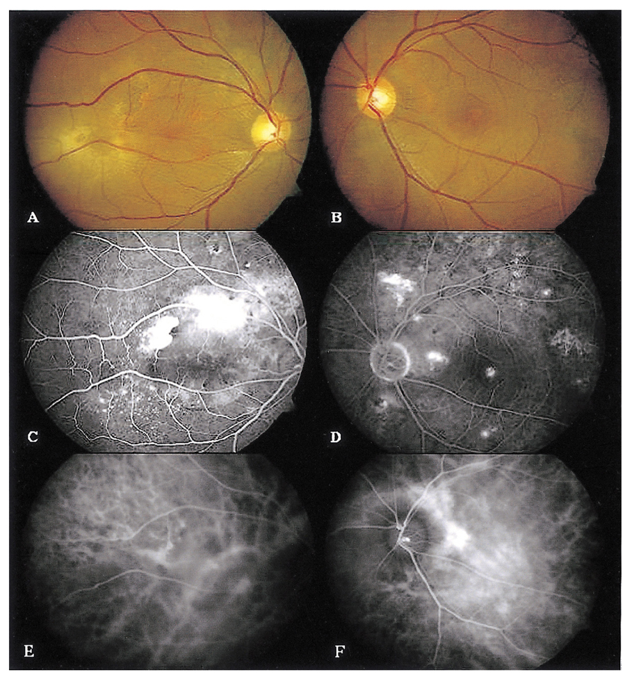
Fig. 1 Color fundus photographs (A, B), fluorescein angiograms (C, D), and indocyanine green angiogram (E, F) before systemic steroid therapy. (A) Right eye. Subretinal fibrosis, retinal fold and pigmented lesion on the posterior pole. (B) Left eye. RPE changes in the macular region and around the vascular arcade. (C) Right eye. Multiple points of dye leakage and pooling into the subretinal space, and variable blockage and staining of areas of subretinal fibrosis. (D) Left eye. Multiple pinpoint leakages. (E) Right eye and (F) left eye, showing multiple choroidal vascular hyperpermeability and focal staining of pigment epithelium.

Fig. 2 Color fundus photographs (A, B), fluorescein angiograms (C, D), and indocyanine green angiogram (E, F) two weeks after systemic steroid therapy. (A) Right eye. Progressed exudative retinal detachment, subretinal fibrosis, retinal fold and pigmented lesion on the posterior pole. (B) Left eye. Multiple localized PEDs. (C) Right eye. Multiple points of dye leakage and pooling into the subretinal space, and variable blockage and staining of areas of subretinal fibrosis. (D) Left eye. Multiple pinpoint leakages. (E) Right eye. (F) Left eye, multiple choroidal vascular hyperpermeability and focal staining of the pigment epithelium.

Fig. 3 Color fundus photographs (A, B), fluorescein angiograms (C, D), and indocyanine green angiogram (E, F) at the time that oral steroid and immunosuppresive treatment were stopped. (A) Right eye. Inferior bullous exudative RD, subretinal fibrotic band, fixed retinal fold, and subtle intraretinal hemorrhages. (B) Left eye. Multiple PEDs and exudative flecks. (C) Right eye. Large blockage of an area of exudative RD, variable blockage and staining of areas of subretinal fibrosis, and leakage and pooling into the subretinal space. (D) Left eye. Multiple pinpoint leakages and poolings. (E) Right eye. (F) left eye. Multiple choroidal vascular hyperpermeability and focal staining of the pigment epithelium.

Fig. 4 B-scan (A, B). showing shifting of subretinal fluid. (A) sitting position. (B) supine position.
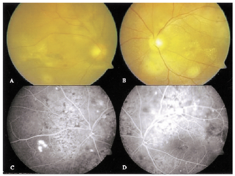
Fig. 5 Color fundus photographs (A, B) and fluorescein angiograms (C, D) six weeks after external drainage of subretinal fluid in the right eye. (A) Right eye. Attached retina with subretinal proliferation. (B) Left eye. Multiple PEDs and exudative flecks. (C) Right eye, Eye absorbed subretinal fluid. (D) Left eye. Multiple points of dye leakage and pooling into the subretinal space.
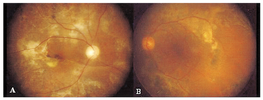
Fig. 6 Color fundus photographs (A, B) after vitrectomy and internal drainage of subretinal fluid of both eyes. (A) Right eye and (B) left eye. Demonstrating an attached retina with atrophic change and subretinal proliferation.
Reference
-
1. Yap EY, Robertson DM. The long-term outcome of central serous chorioretinopathy. Arch Ophthalmol. 1996. 114:689–692.2. Robertson DM. Argon laser photocoagulation treatment in central serous chorioretinopathy. Ophthalmology. 1986. 93:972–974.3. Ficker L, Vafidis G, While A, Leaver P. Long-term follow-up of a prospective trial of argon laser photocoagulation in the treatment of central serous retinopathy. Br J Ophthalmol. 1988. 72:829–834.4. Yoon IS, Do SJ, Im SJ. A case of atypical central serous chorioretinopathy with bullous retinal detachment. J Korean Ophthalmol Soc. 1991. 32:997–1002.5. Kwak HW, Lee MA. Clinical study of atypical central serous chorioretinopathy with bullous retinal detachment. J Korean Ophthalmol Soc. 1994. 35:429–435.6. Ahn DG, Kang SW. The clinical evaluation of atypical idiopathic central serous chorioretinopathy. J Korean Ophthalmol Soc. 2000. 41:691–700.7. Shrma T, Badrinath SS, Gopal L, et al. Subretinal fibrosis and nonrhegmatogenous retinal detachment associated with multifocal central serous chorioretinopathy. Retina. 1998. 18:23–29.8. Otsuka S, Ohba N, Nakao K. A long-term follow-up study of severe variant of central serous chorioretinopathy. Retina. 2002. 22:25–32.9. Bouzas EA, Karadimas P, Pournaras CJ. Central serous chorioretinopathy and glucocorticoids. Surv Ophthalmol. 2002. 47:431–448.10. Gass JDM, Little H. Bilateral bullous exudative retinal detachment complicationg idiopathic central serous chorioretinopathy during systemic corticosteroid therapy. Ophthalmology. 1995. 102:737–747.11. Hooymans JMM. Fibrotic scar formation in central serous chorioretinopathy developed during systemic treatment with corticosteroids. Graefe's Arch Clin Exp Ophthalmol. 1998. 236:876–879.12. Benson WE, Shields JA, Annesley WH, Tasman W. Idiopathic central serous chorioretinopathy with bullous retinal detachment. Ann Ophthalmol. 1980. 12:920–924.13. Mazzuca DE, Benson WE. Central serous chorioretinopathy: Variants. Surv Ophthalmol. 1986. 31:170–174.14. Akiyama K, Kawamura M, Ogata T, Tanaka E. Retinal vascular loss in idiopathic central serous chorioretinopathy with bullous retinal detachment. Ophthalmology. 1987. 94:1605–1609.15. Gass JDM. Bullous retinal detachment: An unsual manifestation of idiopathic centeral serous chorioretinopathy. Am J Ophthalmol. 1973. 75:810–821.16. Schatz H, McDonald R, Johnson RN, et al. Subretinal fibrosis in central serous chorioretinopathy. Ophthalmology. 1995. 102:1077–1088.17. Sahu DK, Namperumalsamy P, Hilton GF, Sousa NF. Bullous variant of idiopathic central serous chorioretinopathy. Br J Ophthalmol. 2000. 84:485–492.18. Bandello F, Incorvaia C, Parmeggiani F, Sebastiani A. Idiopathic multiple serous detachments of the retinal pigment epithelium followed by bilateral central serous chorioretinopathy: a case report. Ophthalmologica. 2000. 214:362–367.19. Lim SS, Chang WH, Kim SD. A case of central serouschorioretinopathy with bullous retinal detachment. J Korean Ophthalmol Soc. 1998. 39:1902–1907.20. Adán A, Corcóstegui B. Surgical management of exudative retinal detachment associated with central serous chorioretinopathy. Ophthalmologica. 2001. 215:74–76.21. Anand R, Tasman WS. Ryan SJ, editor. Nonrhegmatogenous retinal detachment. Retina. 2001. v. 3. St Louis: Mosby;chap. 125.22. Ciardella AP, Guyer DR, Spitznas M, Yannuzzi LA. Ryan SJ, editor. Central serous chorioretinopathy. Retina. v. 2. St Louis: Mosby;chap. 68.23. Kang SE, Kwon HN, Lee HW, et al. A new instrument for drainage or injection of fluid within subretinal space. Retina. 2003. 23:661–666.24. Chen HC, Ho JD, Chen SN. Perfluorocarbon liquid-associated external drainage in the management of central serous chorioretinopathy with bullous serous retinal detachment. Chan Gung Med J. 2003. 26:777–781.
- Full Text Links
-
- Actions
-
Cited
- CITED
-
- Close
- Share
-
- Similar articles
-
- A case of Atypical Central Serous Chorioretinopathy with Bullous Retinal Detachment
- A Case of Central Serous Chorioretinopathy with Bullous Retinal Detachment
- A Case of Bilateral Central Serous Chorioretinopathy in a Chronic Tibial Osteomyelitis Patient
- Measurement and Analysis of Neurosensory Retinal Detachment in Central Serous Chorioretinopathy Using Heidelberg Retina Tomograph
- Central Serous Chorioretinopathy in a Patient with Retinal Macrovessel
